- Title
-
Canonical Wnt signaling through Lef1 is required for hypothalamic neurogenesis
- Authors
- Lee, J.E., Wu, S.F., Goering, L.M., and Dorsky, R.I.
- Source
- Full text @ Development
|
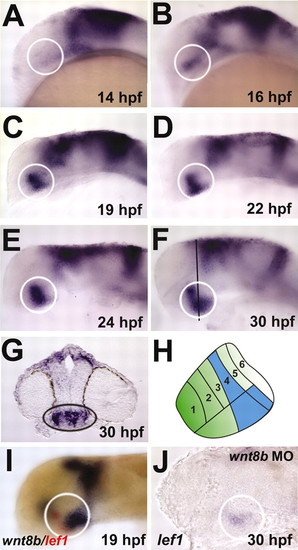
lef1 is expressed in the posterior hypothalamus during embryonic development. Lateral views are shown with anterior towards the left. White circles outline posterior hypothalamus. (A) At 14 hpf, lef1 is strongly expressed in the dorsal midbrain, but does not show specific expression in the developing hypothalamus. (B-F) At 16 hpf, lef1 expression first appears in the presumptive posterior hypothalamus, and this expression is maintained through 30 hpf. After 19 hpf, expression is present in dorsal and ventral regions of the posterior hypothalamus. Black line in F indicates plane of section in G. (G) Transverse section through the posterior hypothalamus (black oval) at 30 hpf, showing lef1 expression in both the medial mitotic cells and the lateral postmitotic cells of the posterior region. (H) Schematic depiction of lef1 expression domain in 30 hpf zebrafish hypothalamus. Numbered regions are based on those of Hauptmann and Gerster (Hauptmann and Gerster, 2000), and lef1 expression is shown in blue. (I) At 19 hpf, wnt8b (blue) and lef1 (red) show overlapping and adjacent expression in the posterior hypothalamus. (J) In wnt8b morphants, lef1 expression is reduced throughout the brain. EXPRESSION / LABELING:
PHENOTYPE:
|
|
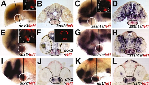
lef1 is co-expressed with proneural and neuronal markers in the posterior hypothalamus. White circles outline posterior hypothalamus and inset panels show lef1 mRNA expression from Fast Red fluorescence. Lines in whole-mount images show plane of corresponding cross-sections on the right. In cross-sections, black ovals outline posterior hypothalamus. (A,B) At 19 hpf, sox3 (blue) is not co-expressed with lef1 (red) in the posterior hypothalamus. (C,D) At 24 hpf, zash1a (blue) is not co-expressed with lef1 (red) in the posterior hypothalamus. (E-H) By 30 hpf, sox3 and zash1a are co-expressed with lef1 in both medial progenitors and lateral postmitotic neurons of the posterior hypothalamus. (I-L) By contrast, dlx2 and isl1 are co-expressed only with lef1 in lateral postmitotic neurons. EXPRESSION / LABELING:
|
|
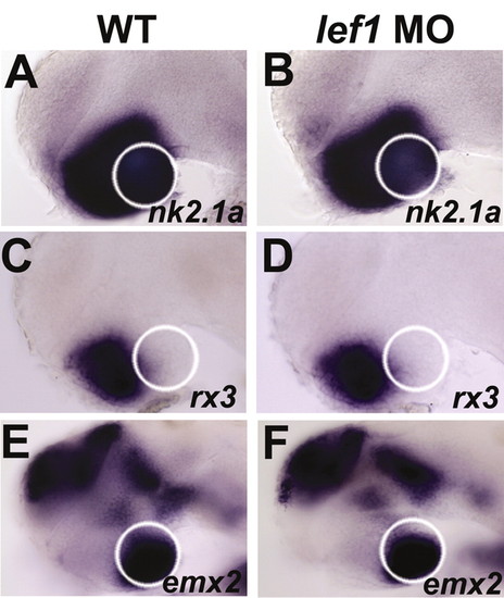
Lef1 is not required for molecular markers of hypothalamus identity or AP patterning. White circles outline posterior hypothalamus. (A,C,E) Uninjected embryos. (B,D,F) Embryos injected with 2 ng of lef1 MO. (A,B) nk2.1a expression in the entire hypothalamus is unaffected in lef1 morphants at 30 hpf. (C,D) rx3 is still expressed in the anterior hypothalamus in lef1 morphants at 30 hpf. (E,F) emx2 is still expressed in the posterior hypothalamus of lef1 morphants at 30 hpf. EXPRESSION / LABELING:
|
|
Lef1 is required for proneural and neuronal gene expression in the posterior hypothalamus. White circles outline posterior hypothalamus in whole-mount views, and black ovals outline posterior hypothalamus in cross-sections. Lines in whole-mount images show plane of corresponding cross-sections. (A-D) Expression of sox3 is absent in the posterior hypothalamus of lef1 morphants at 24 hpf. (E-H) Expression of zash1a is absent in the posterior hypothalamus of lef1 morphants at 28 hpf. (I-P) Expression of dlx2 and isl1 are absent in the posterior hypothalamus of lef1 morphants at 30 hpf. (Q-T) Expression of ngn1 and olig2, which are expressed in the posterior tuberculum, is unaffected in the posterior tuberculum of lef1 morphants. |
|
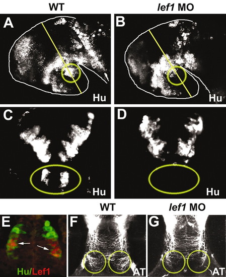
Lef1 is required for neuronal differentiation in the posterior hypothalamus. (A,B) Confocal projections through the hypothalamus of whole-mount 36 hpf brains stained for HuC/D, a pan-neuronal marker. Owing to differences in mounting, neurons outside the hypothalamus may not be visible. (C,D) Cross-sections of 36 hpf brains through the plane marked by the lines in A,B. A specific population of Hu-positive neurons (yellow circles and ovals) is absent in the posterior hypothalamus of lef1 morphants (B,D). (E) Hu (green) is co-expressed with Lef1 protein (red) in a subset of posterior neurons at 36 hpf. (F,G) Acetylated tubulin (AT) staining labels specific axons in the posterior hypothalamus (yellow circles) that are reduced in lef1 morphants at 48 hpf. Confocal projections through the ventral side of the hypothalamus are shown, with anterior towards the top. |
|
Analysis of proliferation and apoptosis in lef1 morphants. (A-L) In lef1 morphants, pH3-positive cells are observed in the posterior hypothalamus at 19, 24 and 30 hpf, indicating that proliferating cells are still present in this region. (A,B,E,F,I,J) Cross-sections through posterior hypothalamus (outlined by dotted circles). (C,D,G,H,K,L) Confocal projections of pH3-stained embryos used for quantitative analysis. The central region outlined by broken lines was defined as posterior hypothalamus, based on morphology in bright-field views. The region farthest to the right was excluded from counts because it does not express lef1 and is unaffected in lef1 morphants (Fig. 4Q-T). (M-P) Apoptotic cells identified by TUNEL staining are absent in the posterior hypothalamus at 19 hpf and 24 hpf in wild-type and lef1 MO-injected embryos (arrowheads). Asterisks indicate apoptotic cells in the dorsal brain of lef1 morphants. |
|
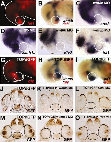
Wnt8b and Lef1 act through the canonical Wnt pathway in the posterior hypothalamus. White circles outline posterior hypothalamus in whole-mount views, and black ovals outline posterior hypothalamus in cross-sections. (A,B) At 30 hpf, wnt8b (blue) and lef1 (red) show overlapping and adjacent expression in the posterior hypothalamus. (C-F) In wnt8b morphants, sox3, zash1a, dlx2 and isl1 expression are specifically reduced in the posterior hypothalamus at 30 hpf. (G-I) A transgenic reporter for ß-catenin-mediated transcription (TOPdGFP) shows mRNA expression in the posterior hypothalamus (red) at 24 hpf. In the posterior hypothalamus, wnt8b expression (blue-H) partially overlaps with gfp, whereas lef1 (blue-I) almost completely overlaps with gfp. (J-O) In TOPdGFP embryos, GFP protein (brown) is expressed in the posterior hypothalamus. At 24 hpf (J-L) and 30 hpf (M-O), this expression is absent in wnt8b and lef1 morphants. |
|
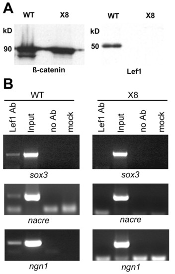
Lef1 associates with the sox3 promoter in vivo. (A) Western blot of whole-embryo lysates probed with anti-β-catenin and Lef1 polyclonal antibodies. The Lef1 band is specifically absent in X8 mutants, whereas the positive control β-catenin band is present. (B) ChIP analysis of whole-embryo lysates shows that the Lef1 antibody can immunoprecipitate DNA fragments containing Lef-binding sites in the upstream regulatory region of sox3, nacre and ngn1. These fragments do not precipitate in controls lacking antibody or chromatin (mock). In lysates from X8 mutants that lack Lef1 protein, the antibody is unable to precipitate these fragments, suggesting that the interaction is specific to Lef1. EXPRESSION / LABELING:
|
|
Injection of a splice-blocking morpholino results in the loss of lef1 mRNA transcripts. (A) Diagram of lef1 gene structure indicating the location of the lef1 MO target at the exon 7 splice donor site. Introns are not to scale. (B) RT-PCR analysis demonstrates that injection of the lef1 MO into zebrafish embryos at the one-cell stage results in the production of aberrantly spliced forms at 12 hpf and the loss of lef1 mRNA after 18 hpf. |
|
Embryos homozygous for the X8 deletion phenocopy Lef1 morphants. (A-C) nk2.1a, rx3 and emx2 expression are unaffected in X8 mutants at 30 hpf. (D-G) sox3, zash1a, dlx2 and isl1 are all absent in the posterior hypothalamus of X8 mutants at 30 hpf. White circles outline the posterior hypothalamus. EXPRESSION / LABELING:
|
|
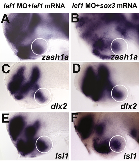
Rescue of lef1 MO phenotypes by lef1 and sox3 mRNA injection. Co-injection of 2 ng lef1 MO with 100 pg lef1 mRNA (A,C,E) or 20 pg sox3 mRNA (B,D,F) rescued the expression of zash1a (A,B), dlx2 (C,D), and isl1 (E,F) in the posterior hypothalamus at 30 hpf. White circles outline the posterior hypothalamus. |

