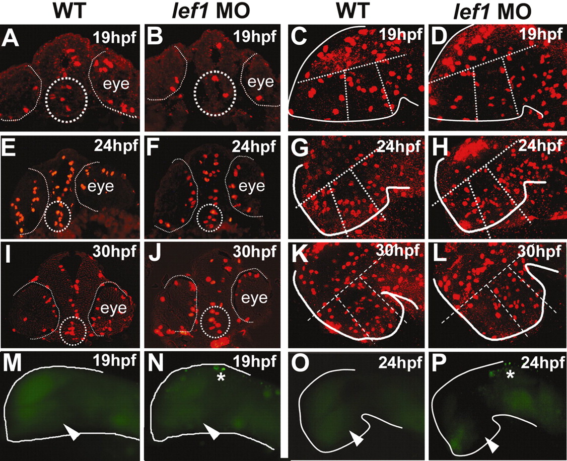
Fig. 6 Analysis of proliferation and apoptosis in lef1 morphants. (A-L) In lef1 morphants, pH3-positive cells are observed in the posterior hypothalamus at 19, 24 and 30 hpf, indicating that proliferating cells are still present in this region. (A,B,E,F,I,J) Cross-sections through posterior hypothalamus (outlined by dotted circles). (C,D,G,H,K,L) Confocal projections of pH3-stained embryos used for quantitative analysis. The central region outlined by broken lines was defined as posterior hypothalamus, based on morphology in bright-field views. The region farthest to the right was excluded from counts because it does not express lef1 and is unaffected in lef1 morphants (Fig. 4Q-T). (M-P) Apoptotic cells identified by TUNEL staining are absent in the posterior hypothalamus at 19 hpf and 24 hpf in wild-type and lef1 MO-injected embryos (arrowheads). Asterisks indicate apoptotic cells in the dorsal brain of lef1 morphants.
Image
Figure Caption
Acknowledgments
This image is the copyrighted work of the attributed author or publisher, and
ZFIN has permission only to display this image to its users.
Additional permissions should be obtained from the applicable author or publisher of the image.
Full text @ Development

