
- Title
-
Photoreceptor regeneration occurs normally in microglia-deficient irf8 mutant zebrafish following acute retinal damage
- Authors
- Song, P., Parsana, D., Singh, R., Pollock, L.M., Anand-Apte, B., Perkins, B.D.
- Source
- Full text @ Sci. Rep.
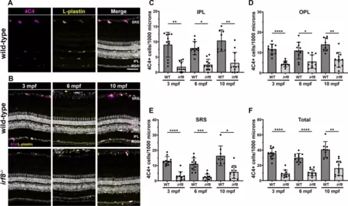
Adult irf8 mutants have a significantly fewer retinal microglia. (A) 4C4 (magenta) and L-plastin (yellow) immunoreactivity in 6 mpf wild-type retinas to label microglia. (B) Immunohistochemistry of 3 mpf, 6 mpf, and 10 mpf wild-type (top row) and irf8 mutants (bottom row) with 4C4 (magenta) and L-plastin (yellow). (C?F) Quantification of 4C4+ cells in the inner plexiform layer (IPL) outer plexiform layer (OPL), subretinal space (SRS), and total retina of wild-type and irf8 mutants with age. Data are plotted as means � SD and p-values were generated by Welch?s ANOVA test with Dunnett?s T3 multiple comparisons test (ONL, SRS, total) or Kruskal?Wallis test with Dunn?s correction for multiple comparisons (INL). *p < 0.05, **p < 0.01, ***p < 0.005, ****p < 0.0001. SRS subretinal space, ONL outer nuclear layer, OPL outer plexiform layer, INL inner nuclear layer, IPL inner plexiform layer, RGC retinal ganglion cell layer. Scale bars, 50 ?m (A,B). |
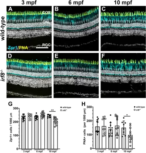
Cone photoreceptors degenerate in 10 mpf adult irf8 mutants. (A?F) Immunohistochemistry with Zpr1 (magenta) and peanut agglutinin (PNA; yellow) on retinal cryosections from wild-type (A?C) and irf8 mutants (D?E) at 3 mpf, 6 mpf, and 10 mpf. (G) Quantification of Zpr1+ cell density in the dorsal retina of wild-type and irf8 mutants with age. (H) Quantification of PNA+ cone outer segments in the dorsal retina of wild-type and irf8 mutants with age. Data are plotted as means � SD and p-values were generated by Welch?s ANOVA test with Dunnett?s T3 multiple comparisons test (Zpr1) or an unpaired t-test (PNA). *p < 0.05, **p < 0.01. COS cone outer segments, ONL outer nuclear layer, INL inner nuclear layer, RGC retinal ganglion cell layer. Scale bar, 50 ?m. |

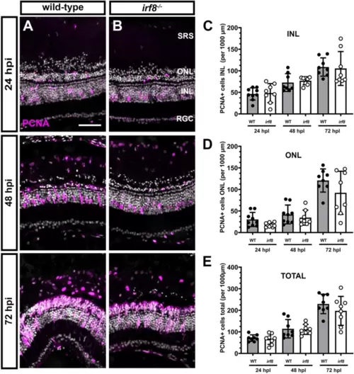
Proliferation of cells in the INL increases in 10 mpf irf8 mutants. (A?F) Immunohistochemistry with anti-PCNA antibodies (magenta) on retinal cryosections from wild-type (A?C) and irf8 mutants (D,E) at 3 mpf, 6 mpf, and 10 mpf. (G?I) Quantification of PCNA+ cell density in the ONL, INL, and total PCNA density of wild-type and irf8 mutants with age. Data are plotted as means � SD and individual points represent data from one retina (n = 8?12). All data were assessed for normal distributions and equal standard deviations. All p-values were generated by unpaired t-tests or unpaired t-tests with Welch?s correction; **p < 0.01. ONL outer nuclear layer, INL inner nuclear layer, RGC retinal ganglion cell layer. Scale bar, 50 ?m. |
Light damage induces proliferation of M�ller glia and generation of neural progenitors in irf8 mutants. (A,B) Immunohistochemistry with anti-PCNA antibodies (magenta) on retinal cryosections from 6 mpf wild-type and irf8 mutants at 24, 48, and 72 h post light injury (hpi). At 24 hpi, proliferating M�ller glia are seen in the INL of both wild-type and irf8 mutants. At 48 hpi, clusters of neurogenic progenitors appear and by 72 hpi these clusters continue to proliferate and have migrated to the ONL of both wild-type and irf8 mutants. (C?E) Quantification of PCNA+ cell density in the INL and ONL and total density of PCNA+ cells in the dorsal retinas of wild-type and irf8 mutants at times after light damage. No statistical difference was found between wild-type and irf8 mutants at any time point. Data are plotted as means � SD and individual points represent data from one retina (n = 10?13). Wild-type and irf8 data were compared by unpaired Welch?s t-tests for individual time points. No statistical difference was observed between any groups. SRS subretinal space, ONL outer nuclear layer, INL inner nuclear layer, RGC retinal ganglion cell layer. Scale bar, 50 ?m. |
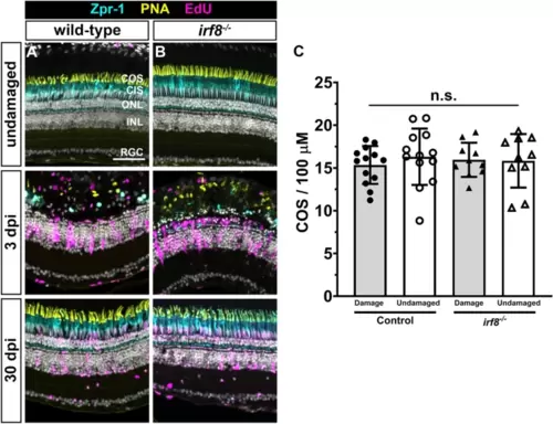
Photoreceptors regenerate in irf8 mutants following acute light damage. (A,B) Immunohistochemistry with Zpr1 (cyan), PNA (yellow), and EdU (magenta) on retinal cryosections from undamaged wild-type and irf8 mutants (top row) and at 3 days post injury (dpi; middle row) or 30 dpi (bottom row). Light damage fully ablated cones in the central region of the dorsal retina at 3 dpi and photoreceptors regenerated by 30 dpi. (C) Quantification of cone outer segment density in damaged and undamaged areas of the retina. Data are plotted as means � SD and individual points represent data from one retina (n = 8?9). Data were analyzed by Welch?s ANOVA test with Dunnett T3 correction for multiple comparisons. No statistical difference was observed between any groups. COS cone outer segments, CIS cone inner segments, ONL outer nuclear layer, INL inner nuclear layer, RGC retinal ganglion cell layer. Scale bar, 50 ?m. |
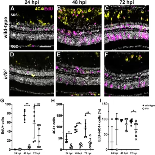
Light damage does not induce microglia proliferation in irf8 mutants. (A?F) Immunohistochemistry with EdU (magenta) to label proliferating cells and 4C4 (yellow) to label microglia/macrophages on retinal cryosections from wild-type and irf8 mutants at 24, 48, and 72 h post injury (hpi). (G) Quantification of EdU+ cells in the SRS at times after light damage. (H) Quantification of 4C4+ cells in the SRS at times after light damage. (I) The ratio of Edu+/4C4+ cells within the SRS was calculated for each time point after light damage. SRS subretinal space, ONL outer nuclear layer, INL inner nuclear layer, RGC retinal ganglion cell layer. Scale bar, 50 ?m. |
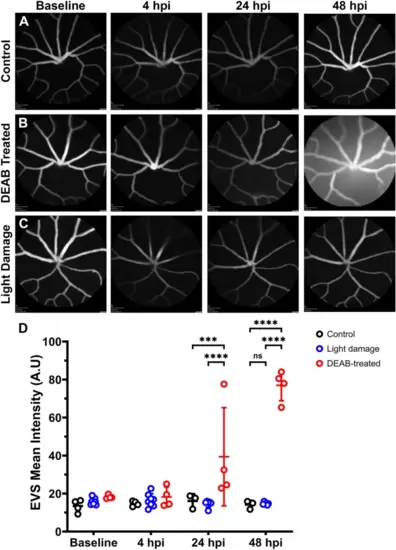
Light damage does not disrupt the blood-retinal-barrier. (A?C) Longitudinal confocal scanning laser ophthalmoscopy (cSLO) images from 6 mpf Tg(l-fabp:DBP-eGFP) zebrafish before and at 4?48 h after treatment with DMSO, DEAB, or with light damage. (D) Quantification of extravascular space (EVS) mean signal intensity determined from cSLO images at different time points after treatment. Data were analyzed with a two-way ANOVA with Tukey?s correction for multiple comparisons. ***p < 0.0005; ****p < 0.0001. |
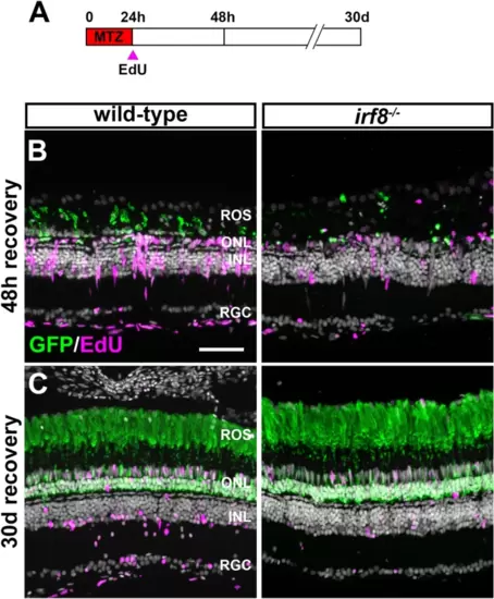
Regeneration after metronidazole-induced rod ablation in irf8;Tg(zOPS:ntrB-EGFP)gm500 animals. (A) Schematic illustrating experimental paradigm and treatment regimen. (B,C) Immunohistochemistry with anti-GFP antibodies (green) and EdU (magenta) on retinal cryosections from wild-type and irf8 mutants at 48 h post MTZ treatment or 30 days post MTZ treatment. MTZ treatment fully ablated rod photoreceptors and induced M�ller glia proliferation at 48 h post treatment and rod fully regenerated by 30 dpi. ROS rod outer segments, ONL outer nuclear layer, INL inner nuclear layer, RGC retinal ganglion cell layer. Scale bar, 50 ?m. |
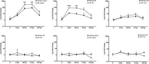
Dynamics of inflammatory signaling following light damage in 6 mpf irf8 mutants and wild-type siblings. The change in gene expression of inflammatory genes following light damage is plotted as a function of time after damage to wild-type or irf8 mutants. Expression is represented as the log fold change calculated using the ddCt method and compared against gene expression in undamaged retinas. ***p < 0.0005; **p < 0.01; *p < 0.05. |