
|
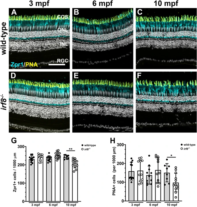
Fig. 2 Cone photoreceptors degenerate in 10 mpf adult irf8 mutants. (A?F) Immunohistochemistry with Zpr1 (magenta) and peanut agglutinin (PNA; yellow) on retinal cryosections from wild-type (A?C) and irf8 mutants (D?E) at 3 mpf, 6 mpf, and 10 mpf. (G) Quantification of Zpr1+ cell density in the dorsal retina of wild-type and irf8 mutants with age. (H) Quantification of PNA+ cone outer segments in the dorsal retina of wild-type and irf8 mutants with age. Data are plotted as means � SD and p-values were generated by Welch?s ANOVA test with Dunnett?s T3 multiple comparisons test (Zpr1) or an unpaired t-test (PNA). *p < 0.05, **p < 0.01. COS cone outer segments, ONL outer nuclear layer, INL inner nuclear layer, RGC retinal ganglion cell layer. Scale bar, 50 ?m.