
|
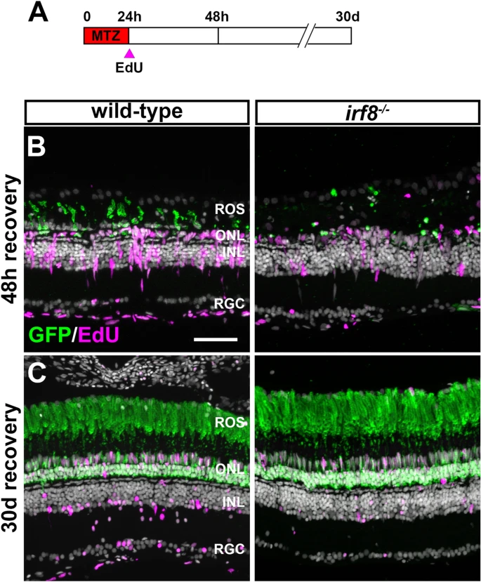
Fig. 8 Regeneration after metronidazole-induced rod ablation in irf8;Tg(zOPS:ntrB-EGFP)gm500 animals. (A) Schematic illustrating experimental paradigm and treatment regimen. (B,C) Immunohistochemistry with anti-GFP antibodies (green) and EdU (magenta) on retinal cryosections from wild-type and irf8 mutants at 48 h post MTZ treatment or 30 days post MTZ treatment. MTZ treatment fully ablated rod photoreceptors and induced M�ller glia proliferation at 48 h post treatment and rod fully regenerated by 30 dpi. ROS rod outer segments, ONL outer nuclear layer, INL inner nuclear layer, RGC retinal ganglion cell layer. Scale bar, 50 ?m.