
Fig. 1
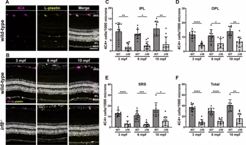
Adult irf8 mutants have a significantly fewer retinal microglia. (A) 4C4 (magenta) and L-plastin (yellow) immunoreactivity in 6 mpf wild-type retinas to label microglia. (B) Immunohistochemistry of 3 mpf, 6 mpf, and 10 mpf wild-type (top row) and irf8 mutants (bottom row) with 4C4 (magenta) and L-plastin (yellow). (C?F) Quantification of 4C4+ cells in the inner plexiform layer (IPL) outer plexiform layer (OPL), subretinal space (SRS), and total retina of wild-type and irf8 mutants with age. Data are plotted as means � SD and p-values were generated by Welch?s ANOVA test with Dunnett?s T3 multiple comparisons test (ONL, SRS, total) or Kruskal?Wallis test with Dunn?s correction for multiple comparisons (INL). *p < 0.05, **p < 0.01, ***p < 0.005, ****p < 0.0001. SRS subretinal space, ONL outer nuclear layer, OPL outer plexiform layer, INL inner nuclear layer, IPL inner plexiform layer, RGC retinal ganglion cell layer. Scale bars, 50 ?m (A,B). |