
Fig. 4
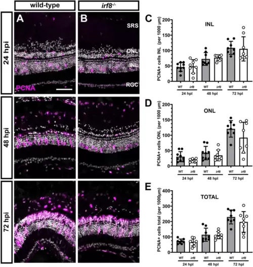
Light damage induces proliferation of M�ller glia and generation of neural progenitors in irf8 mutants. (A,B) Immunohistochemistry with anti-PCNA antibodies (magenta) on retinal cryosections from 6 mpf wild-type and irf8 mutants at 24, 48, and 72 h post light injury (hpi). At 24 hpi, proliferating M�ller glia are seen in the INL of both wild-type and irf8 mutants. At 48 hpi, clusters of neurogenic progenitors appear and by 72 hpi these clusters continue to proliferate and have migrated to the ONL of both wild-type and irf8 mutants. (C?E) Quantification of PCNA+ cell density in the INL and ONL and total density of PCNA+ cells in the dorsal retinas of wild-type and irf8 mutants at times after light damage. No statistical difference was found between wild-type and irf8 mutants at any time point. Data are plotted as means � SD and individual points represent data from one retina (n = 10?13). Wild-type and irf8 data were compared by unpaired Welch?s t-tests for individual time points. No statistical difference was observed between any groups. SRS subretinal space, ONL outer nuclear layer, INL inner nuclear layer, RGC retinal ganglion cell layer. Scale bar, 50 ?m. |