
|
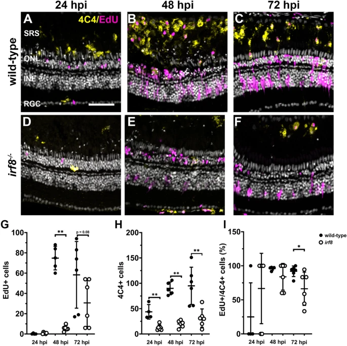
Fig. 6 Light damage does not induce microglia proliferation in irf8 mutants. (A?F) Immunohistochemistry with EdU (magenta) to label proliferating cells and 4C4 (yellow) to label microglia/macrophages on retinal cryosections from wild-type and irf8 mutants at 24, 48, and 72 h post injury (hpi). (G) Quantification of EdU+ cells in the SRS at times after light damage. (H) Quantification of 4C4+ cells in the SRS at times after light damage. (I) The ratio of Edu+/4C4+ cells within the SRS was calculated for each time point after light damage. SRS subretinal space, ONL outer nuclear layer, INL inner nuclear layer, RGC retinal ganglion cell layer. Scale bar, 50 ?m.