
|
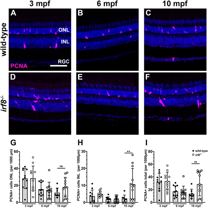
Fig. 3 Proliferation of cells in the INL increases in 10 mpf irf8 mutants. (A?F) Immunohistochemistry with anti-PCNA antibodies (magenta) on retinal cryosections from wild-type (A?C) and irf8 mutants (D,E) at 3 mpf, 6 mpf, and 10 mpf. (G?I) Quantification of PCNA+ cell density in the ONL, INL, and total PCNA density of wild-type and irf8 mutants with age. Data are plotted as means � SD and individual points represent data from one retina (n = 8?12). All data were assessed for normal distributions and equal standard deviations. All p-values were generated by unpaired t-tests or unpaired t-tests with Welch?s correction; **p < 0.01. ONL outer nuclear layer, INL inner nuclear layer, RGC retinal ganglion cell layer. Scale bar, 50 ?m.