
|
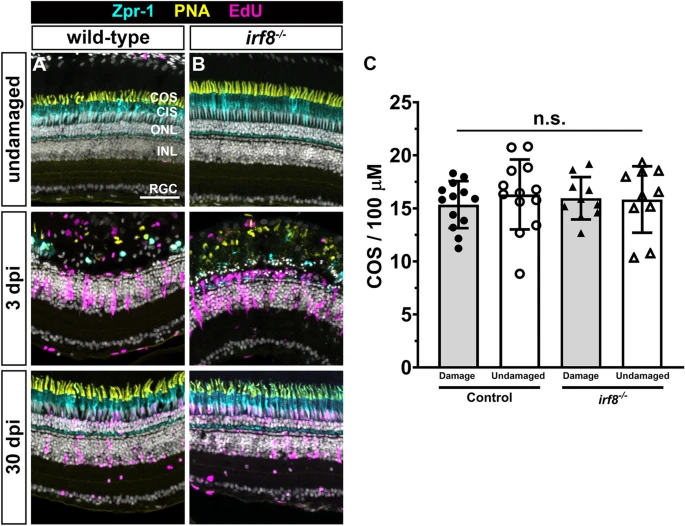
Fig. 5 Photoreceptors regenerate in irf8 mutants following acute light damage. (A,B) Immunohistochemistry with Zpr1 (cyan), PNA (yellow), and EdU (magenta) on retinal cryosections from undamaged wild-type and irf8 mutants (top row) and at 3 days post injury (dpi; middle row) or 30 dpi (bottom row). Light damage fully ablated cones in the central region of the dorsal retina at 3 dpi and photoreceptors regenerated by 30 dpi. (C) Quantification of cone outer segment density in damaged and undamaged areas of the retina. Data are plotted as means � SD and individual points represent data from one retina (n = 8?9). Data were analyzed by Welch?s ANOVA test with Dunnett T3 correction for multiple comparisons. No statistical difference was observed between any groups. COS cone outer segments, CIS cone inner segments, ONL outer nuclear layer, INL inner nuclear layer, RGC retinal ganglion cell layer. Scale bar, 50 ?m.