
Image
|
Figure Caption
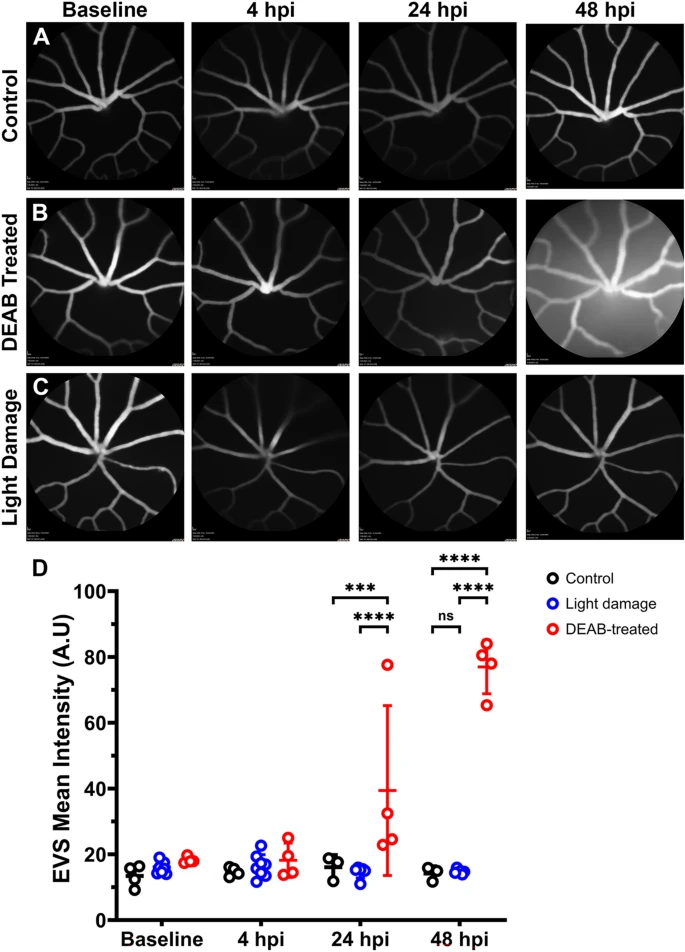
Fig. 7 Light damage does not disrupt the blood-retinal-barrier. (A?C) Longitudinal confocal scanning laser ophthalmoscopy (cSLO) images from 6 mpf Tg(l-fabp:DBP-eGFP) zebrafish before and at 4?48 h after treatment with DMSO, DEAB, or with light damage. (D) Quantification of extravascular space (EVS) mean signal intensity determined from cSLO images at different time points after treatment. Data were analyzed with a two-way ANOVA with Tukey?s correction for multiple comparisons. ***p < 0.0005; ****p < 0.0001.
Acknowledgments
This image is the copyrighted work of the attributed author or publisher, and
ZFIN has permission only to display this image to its users.
Additional permissions should be obtained from the applicable author or publisher of the image.
Full text @ Sci. Rep.