- Title
-
The LIM-homeodomain transcription factor Islet2a promotes angioblast migration
- Authors
- Lamont, R.E., Wu, C.Y., Ryu, J.R., Vu, W., Davari, P., Sobering, R.E., Kennedy, R.M., Munsie, N.M., Childs, S.J.
- Source
- Full text @ Dev. Biol.
|
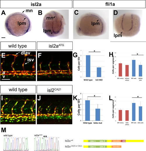
Isl2a is expressed in the lateral posterior mesoderm and promotes organized angioblast migration. In lateral (A) and dorsal posterior (B) views at 12 S, isl2a is expressed in motoneuron cell bodies (mn) and in the lateral posterior mesoderm (lpm). (C-D) In lateral (C) and dorsal posterior (D) views at 12 S, fli1a is expressed in the lateral posterior mesoderm (lpm). Confocal microscopy of Tg(fli1a: neGFP)y7(kdrl:mCherry)ci5 embryos showing a lateral view of the trunk of wild type (E) as compared to isl2aATG morphant (F), and sibling (I) as compared to isl2aCA21 mutant (J) highlighting endothelial cytoplasm (red) and nuclei (green). The number of cells in ISAs were enumerated in morphant (G) and mutant (K) embryos. The number of cells in the artery or vein was enumerated in morphant (H) and mutant (L) embryos. (M) Sequence of isl2aca21 mutants showing a 2 bp insertion leading to a truncation of the protein before the homeodomain sequence. Scale bars represent 100 Ám. EXPRESSION / LABELING:
PHENOTYPE:
|
|
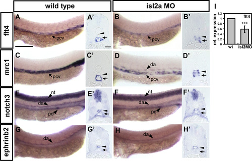
Isl2 modulates vein cell marker expression. Compared to wild type controls (A,C,E,G), expression of the venous markers flt4 (B) and mrc1 (D) is reduced in the trunk of isl2a morphants at 24 hpf, while there is no obvious change in the expression of arterial markers notch3 (F) and ephrinb2 (H). (A?-H?) are cross sections of embryos in A-H. (I) There is a 41% reduction in flt4 expression by qPCR (p<0.0001 by student's t-test) in isl2a morphants. Scale bars represent 200 Ám in A-H and 30 Ám in cross-sections A?-H?. Abbreviations: posterior cardinal vein (pcv), dorsal aorta (da), neural tube (nt) and pronephric ducts (pd). |
|
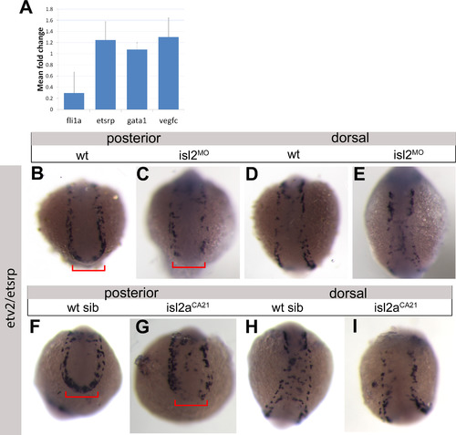
Loss of Isl2a interferes with angioblast migration and fli1a expression. (A) qPCR expression of angioblast genes. (B-I) Expression of etsrp/etv2 in wild type (B,D,F,H), isl2MO (C,E) or isl2aca21 mutants (G,I) as seen in dorsal or posterior views. Red bracket denotes expression at posterior of embryo. EXPRESSION / LABELING:
PHENOTYPE:
|
|
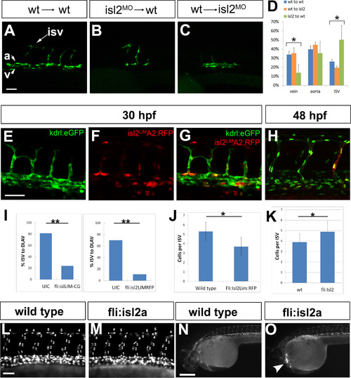
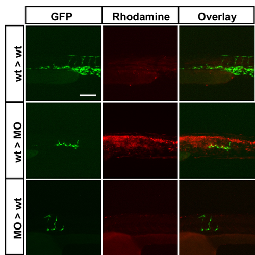
isl2a is required in angioblasts. (A?D) Tg(kdrl:GFP)la116 cells derived from Tg(kdrl: GFP)la116 embryos were transplanted into non-transgenic wild type (wt) or isl2a morphant (MO) recipient embryos. Cells transplanted from wild type donors into wild type recipients (A; blue bars; wt>wt) contribute to the artery, vein and ISAs. Cells from isl2aATG MO injected donor embryos transplanted into wild type recipient embryos (B; MO>wt) contribute less to the vein (green bars; J,L; p<0.02) while wild type cells transplanted into isl2aATG morphant recipient embryos (C; orange bars; wt>MO) do not differ statistically from wild type>wild type transplants in their contribution to the artery, vein and ISAs. (E?J) Expression of isl2aLim in wild type embryos results in stalled ISAs at 30 hpf (E: kdrl: GFP, F: Isl2LimA2TagRFP, G: overlay) and fewer cells per ISA at 48 hpf (H: green Tg(fli: neGFP)y7, red Isl2LimA2TagRFP). (I) ISA stalling is similar at 30 hpf in Isl2Lim injected embryos in two different vectors with different tagging systems. (J) The number of cells per ISA is significantly reduced in dominant negative injected embryos. (K). The number of cells per ISA is significantly increased with angioblast expression of isl2a. (L?O) Effect of upregulation of isl2a in angioblasts. The number of cells forming each ISA was counted in wild type control (L) and fli1a:isl2a (M) overexpressing embryos at 30 hpf. (N,O) Morphology of fli1a:isl2a overexpressing embryos is not distinguishable from wild type embryos and success of transgenesis can be verified expression of cmlc (cardiomyocyte light chain) driven GFP from the vector backbone (arrowhead) in the heart. Scale bars represent 50 Ám for A-H, L?M, and H 100 Ám for M-O. |
|
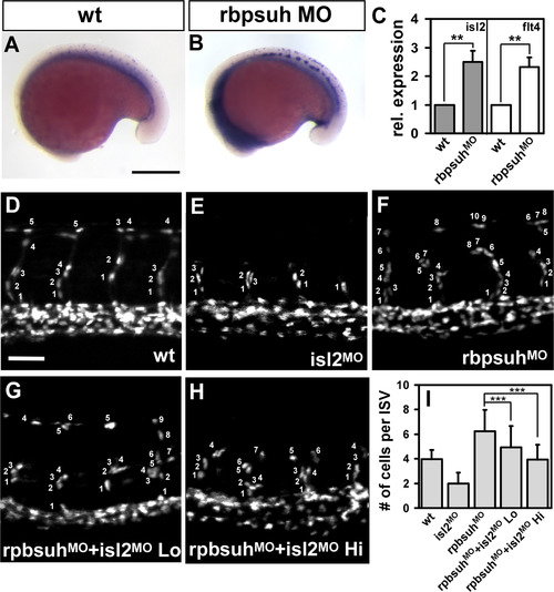
Islet2a is negatively regulated by Notch signaling. (A?C) isl2a expression is strongly upregulated in rbpsuh morphants (B) as compared to control embryos (A). (C) Relative expression levels of isl2a and flt4 was increased by 2.5-fold and 2.3-fold, respectively, in rbpsuh morphants. (D?H) Representative photomicrographs showing the number of nuclei per ISA at 30 hpf in (D) wild type embryos, (E) Isl2ATG MO, (F) Rbpsuh MO, (G) Rbpsuh MO with Isl2ATG MO Lo (5.1 ng), (H) Rbpsuh MO with Isl2ATG MO Hi (12.1 ng). (I) Quantitation of the average number of cells per ISA in single and double morphants. ** refers to p<0.005, and *** refers to p<0.0001 by an unpaired student's t-test. Scale bars represent 100 Ám in A, B and 50 Ám in D?H. PHENOTYPE:
|
|
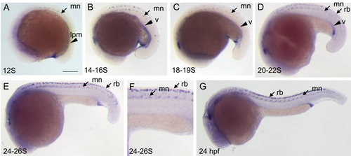
Spatiotemporal expression of isl2 during development A) isl2 expression is first observed at the 12S stage in the lateral posterior mesoderm (lpm) and motoneurons of the trunk (mn). B-C) At both 14-16S and 18-19S, isl2 expression continues in the ventral lateral trunk (v). D) At 20-22S, expression of isl2 is now seen in the Rohon Beard cells of the trunk (rb), as well as motoneurons and the ventral medial trunk. E) Expression in Rohon Beard and motoneurons increases by 24-26S, but expression of isl2 in the ventral trunk is diminished (F, enlargement). G) At 24hpf, expression is no longer seen in the ventral trunk. Scale bar for all panels (except F, which is an enlargement of E) represents 100 ?m. ? EXPRESSION / LABELING:
|
|
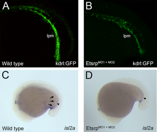
Knockdown of etsrp (etv2) and expression of isl2a A-B) Expression of the vascular endothelial transgenic marker Tg(kdrl:GFP)la116 is reduced by morpholino knockdown of etsrp. C-D) Expression of isl2a is reduced with morpholino knockdown of etsrp (arrows mark lateral plate expression which is strongly reduced with etsrp loss, arrowhead marks midline expression). |
|
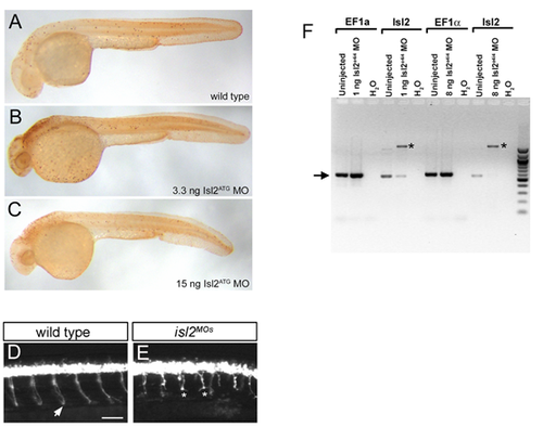
Morpholino controls (A-C) Isl2aTG or e4i4 injected embryos do not show enhanced cell death. TUNEL labeling was used to detect apoptotic cells in wild type, and isl2ATG morphants at two different doses. Although some neural death was observed, cell death in vascular regions was not elevated over that observed in wild type controls. (D-E) Isl2ATG MO injected embryos show motoneuron stalling defects as previously demonstrated: Isl2ATG MO was injected into Tg(mnx1:GFP)ml2 embryos expressing GFP in primary motoneurons. At 24 hpf wild type embryos show normal growth of the caudal primary motoneurons with axons that extend from the spinal cord to the ventral aspect of the embryo (arrow), Embryos injected with isl2ATG MO show stalled axons with increased branching (asterisk). Scale bar represents 50 µm. (F) Knockdown efficiency of isl2 morpholino cDNA from uninjected controls or Isl2 morphants (injected with either 1 ng or 8 ng morpholino) underwent PCR with primers for the housekeeping control gene (EF1?), or for isl2. In morphants injected with isl2e4i4, EF1? levels are unchanged while the amount of wild-type isl2 product is diminished (arrow) and a new higher molecular weight band appears (*), representing mis-splicing caused by the morpholino. Injection of 1 ng of morpholino causes partial mis-splicing, while mis-splicing is almost complete with 8 ng of morpholino. The predicted product size for wild type Isl2 is 547bp. We found a 1663bp mis-spliced band in morphants corresponding to the inclusion of an intron in the mRNA, as detected by sequencing. |
|
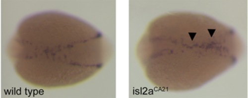
Expression of fli1a in wild type and isl2a mutants shows disorganization of isl2a angioblasts as they migrate to the midline. A decrease to 0.3 expression level of fli1a was detected by qPCR, but is not visible when using the less quantitative in situ hybridization method. |
|
Rhodamine dextran images of representative transplanted embryos. First column: Tg(kdrl:eGFP)la116 donor angioblasts (green) were placed in non-transgenic recipient embryos. Middle column: Rhodamine dextran was included in the transplanted cells to label all transplanted cells (i.e. non-endothelial cells). Signal from rhodamine dextran is shown corresponding to the Tg(kdrl:eGFP)la116 image. Right column: A merged photograph is shown. Scale bar represents 50 µm. |
|
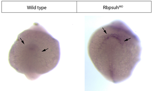
Posterior views of isl2a expression in wild type and rbpsuh morphant embryos Isl2a expression was assayed at 12S in uninjected and rbpsuhMO injected embryos by in situ hybridization. Arrows point to expression (or lack of expression) of Isl2a in the posterior lateral mesoderm. |
|
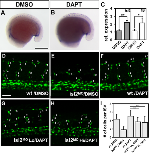
Loss of isl2 compensates for increased ISV angioblast number caused by loss of notch signaling by DAPT (A-C) isl2 expression is upregulated after treatment with DAPT (B) as compared to untreated control embryos (A). (C) Relative expression levels of isl2 and flt4 were quantitated by qPCR. Expression of isl2 and flt4 was increased by 2.2-fold in DAPT-treated embryos. (D-H) Representative embryos showing the number of nuclei per ISV at 30 hpf. (D) Wild type control embryos treated with DMSO, (E) isl2ATG MO, (F) DAPT treatment, (G), DAPT and isl2ATG MO Lo, and (H) DAPT with isl2ATG MO Hi. (I) Quantitation of the average number of cells in single ISVs in DMSO treated control embryos (4.3 ▒ 1.1), isl2ATG morphants (1.8 ▒ 0.7), DAPT treated embryos (5.0 ▒ 1.4), DAPT treatment with isl2ATG Lo (3.7 ▒ 1.0), or DAPT treatment with isl2ATG MO Hi (3.6 ▒ 1.4) (* refers to p< 0.05, ** refers to p<0.005, and *** refers to p<0.0001 by an unpaired student's t-test. Scale bars represent 100 µm in A, B and 50 µm in D-H. |
|
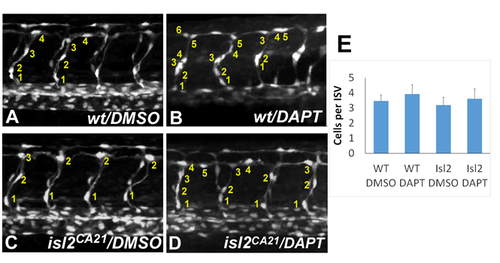
isl2a deficiency compensates for increased ISV cell number caused by loss of notch signaling by DAPT (A-D) Representative embryos showing the number of nuclei per ISV at 30-32 hpf. Wild type control embryos treated with DMSO (A) or DAPT (B), and isl2CA21 mutant embryos treated with DMSO (C) or DAPT (D). (E) Average number of cells per ISV under all four conditions. |
|
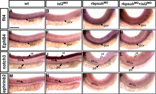
Loss of isl2 compensates for increased venous markers caused by loss of notch signaling in rbpsuh morphants. (A-P) Representative images showing the expression of arterial and venous markers in wild type, isl2ATG MO, rbpsuhMO and rbpsuhMO + isl2ATG MO at 24 hpf. Compared to wild type controls (A, E, I, M), expression of the venous markers flt4 (B) and ephB4 (F) is reduced in the trunk of isl2 morphants while there is no obvious change in the expression of arterial markers notch3 (I) and ephrinb2 (N). In rbpsuhMO, expression of the venous markers flt4 (C) and ephB4 (G) is increased and there is a decrease in the expression of arterial markers notch3 (K) and ephrinb2 (O). In rbpsuhMO + isl2ATG MO double knockdown expression of venous markers (D, H) is similar to wild type; however, the level of arterial markers (L, P) remains the same as rbpsuhMO. Scale bars are 50 m in A-P. |

ZFIN is incorporating published figure images and captions as part of an ongoing project. Figures from some publications have not yet been curated, or are not available for display because of copyright restrictions. PHENOTYPE:
|

Unillustrated author statements |
Reprinted from Developmental Biology, 414, Lamont, R.E., Wu, C.Y., Ryu, J.R., Vu, W., Davari, P., Sobering, R.E., Kennedy, R.M., Munsie, N.M., Childs, S.J., The LIM-homeodomain transcription factor Islet2a promotes angioblast migration, 181-92, Copyright (2016) with permission from Elsevier. Full text @ Dev. Biol.

