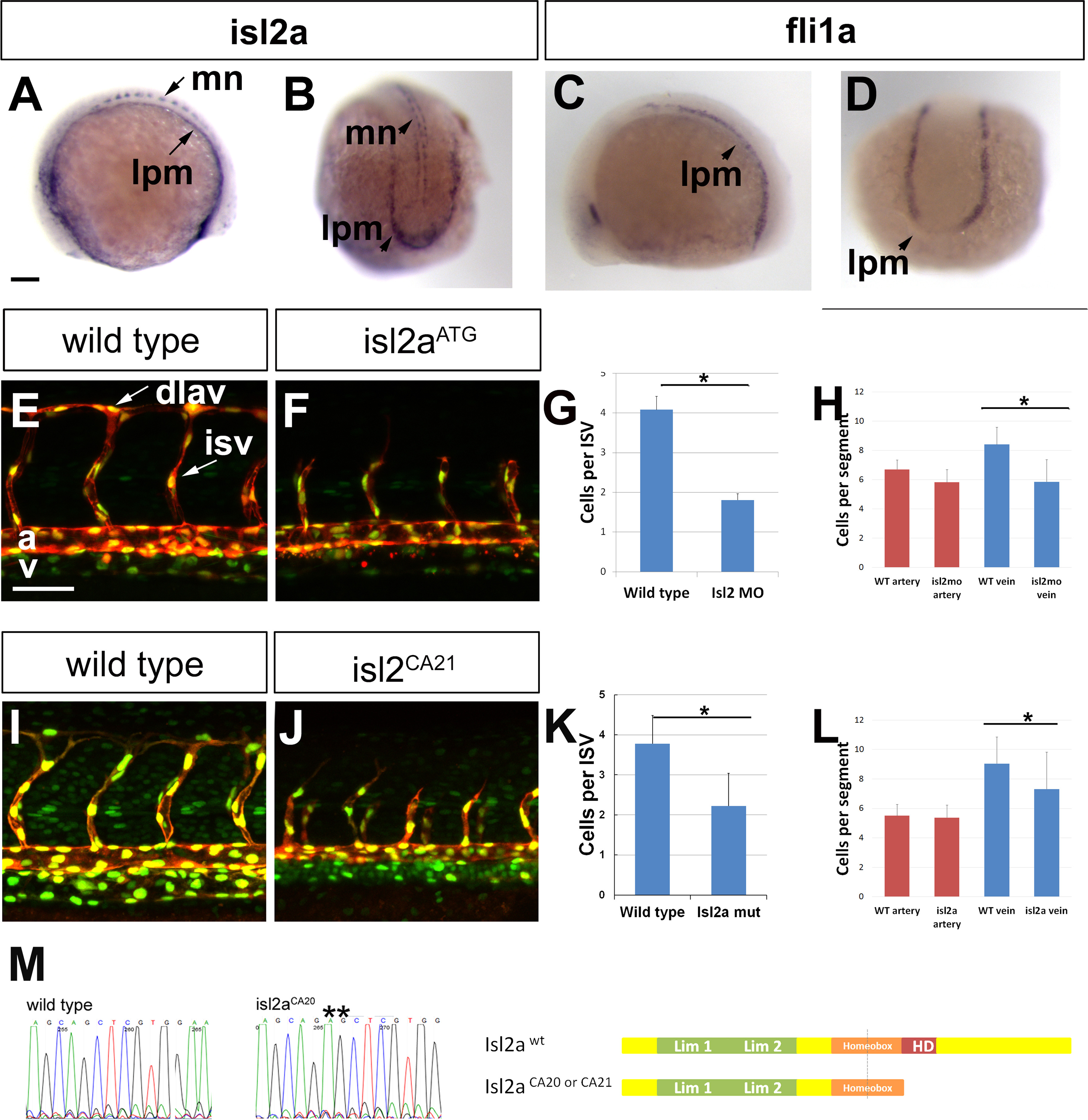
Fig. 1
Isl2a is expressed in the lateral posterior mesoderm and promotes organized angioblast migration. In lateral (A) and dorsal posterior (B) views at 12 S, isl2a is expressed in motoneuron cell bodies (mn) and in the lateral posterior mesoderm (lpm). (C-D) In lateral (C) and dorsal posterior (D) views at 12 S, fli1a is expressed in the lateral posterior mesoderm (lpm). Confocal microscopy of Tg(fli1a: neGFP)y7(kdrl:mCherry)ci5 embryos showing a lateral view of the trunk of wild type (E) as compared to isl2aATG morphant (F), and sibling (I) as compared to isl2aCA21 mutant (J) highlighting endothelial cytoplasm (red) and nuclei (green). The number of cells in ISAs were enumerated in morphant (G) and mutant (K) embryos. The number of cells in the artery or vein was enumerated in morphant (H) and mutant (L) embryos. (M) Sequence of isl2aca21 mutants showing a 2 bp insertion leading to a truncation of the protein before the homeodomain sequence. Scale bars represent 100 Ám.
Reprinted from Developmental Biology, 414, Lamont, R.E., Wu, C.Y., Ryu, J.R., Vu, W., Davari, P., Sobering, R.E., Kennedy, R.M., Munsie, N.M., Childs, S.J., The LIM-homeodomain transcription factor Islet2a promotes angioblast migration, 181-92, Copyright (2016) with permission from Elsevier. Full text @ Dev. Biol.

