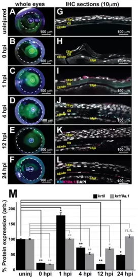
Keratin intermediate filament proteins 8 and 18a.1 are expressed at various time points during postnatal corneal wound healing. A zebrafish adult ocular injury model was established to achieve extensive corneal injury consisting of epithelial debridement, removal of Bowman?s membrane, and excavation of the anterior to mid stroma. (A?F) Fluorescein tracing to distinguish injured corneal surfaces shows the complete healing of the injured eye by 24 h post injury (hpi). The dashed and solid circles highlight the iris and pupil, respectively, in the zebrafish eye. (G?L) Immunohistochemical analysis of thin (10 ?m) sections shows that the corneal stroma regenerated within 1 h following mechanical injury, and the expression of both K8 and K8a.1 in neural crest-derived tissues (stroma and endothelium) was detected in the uninjured (uninj) cornea and during ocular wound healing. CEpi, corneal epithelium; CS, corneal stroma; CEndo, corneal endothelium. (M) Quantitative analysis of K8 and K18a.1 expression shows differential expression patterns during corneal wound healing. *, p-value < 0.05; **, p-value < 0.01; n.s., not significant.
|
