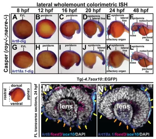
Krt8/krt18a.1 are expressed in the ocular and periocular neural crest during early development. Wholemount in situ hybridization in Casper (roy-/-; nacre-/-) zebrafish embryos during early development at 8, 12, 16, 20, 24, and 48 hpf. K8 and K18a.1 gene expression was detected using a colorimetric assay (Vector Blue Substrate Kit, Vector Laboratories) that is both chromogenic (blue) and fluorescent (Cy5). The sections were mounted in a media containing DAPI (gray). Lateral brightfield wholemount images show krt8 (A–F) and krt18a.1 (G–L) expression initiating dorsally along the neural plate border and enveloping layer (EVL) at 8 hpf (A,G) then dorsoposteriorally in the embryonic epithelium and ventrally in the ocular and craniofacial regions (B–E,H–K), with apparent expression in the ocular anterior segment and facial mesenchyme (blue arrows, (F,L)) by 48 hpf. Fluorescent double in situ hybridization for krt8 (M) or krt18a.1 (N) (Cy5/dark blue) and foxd3 (Texas Red/magenta) expression, followed by immunohistochemistry for sox10-GFP (α-GFP/light blue) was performed on transverse cephalic sections. The black dashed line (lower left insert) indicates the orientation of the plane of section, which passes perpendicular to the spinal column and extends in the rostral-caudal direction. Fluorescent confocal microscopy revealed krt8/krt18a.1 expression in the neural crest-derived ocular anterior segment (primordial cornea, orange arrows) and periocular mesenchyme (yellow arrows) of zebrafish embryos at 24 hpf.
|
