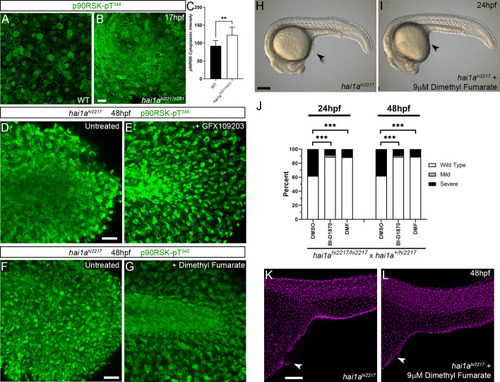
Figure 10—figure supplement 1.

RSK inhibitors rescue the <italic toggle='yes'>hai1a</italic> phenotype.

(A, B) Lateral projected confocal images of the yolk surface of 17hpf WT (A) and hai1ahi2217/ti251 (B) embryos immunostained for p90RSK (Phospho-Thr348). (C) Quantification of immunofluorescent intensity of cytoplasmic levels of p90RSK (Phospho-Thr348) in cells over the yolk of 17hpf WT and hai1ahi2217/ti251. Measurements were limited to keratinocyte cytoplasm by reading at sites between TP63-positive nuclei. n = 9 (three cells from three embryos each); Mann–Whitney test; **p<0.01. (D–G) Lateral projected confocal images of the tails of 48hpf hai1ahi2217 embryos, untreated (D, F) or treated with GFX109203 to inhibit PKC (E) or dimethyl fumarate (DMF) (G) and then immunostained for p90RSK (Phospho-Thr348). Levels of cytoplasmic p90RSK-Thr348 are reduced by both inhibitors. (H, I) Lateral DIC images of hai1ahi2217 embryos at 24hpf either untreated (H) or treated with 9 µM DMF (I). Locations of epidermal aggregates and loss of tail fin morphology in hai1a mutants, and their rescue by RSK inhibitor treatment are indicated by arrowheads. (J) Proportions of epidermal phenotypes from hai1ahi2217/hi2217 × hai1a+/hi2217 determined at 24hpf and 48hpf following treatments with DMSO, BI-D1870, or DMF. n = 100; Chi-squared test; ***p<0.001. (K, L) Lateral projected confocal images of trunks of 48hpf hai1ahi2217 embryos, untreated (K) or treated with 9 µM DMF (L), and then fluorescently immunostained for TP63. Scale bars: (A) = 200 µm; (D) = 100 µm.

Expression Data

Expression Detail
Antibody Labeling
Phenotype Data

Phenotype Detail
Acknowledgments
This image is the copyrighted work of the attributed author or publisher, and ZFIN has permission only to display this image to its users. Additional permissions should be obtained from the applicable author or publisher of the image. Full text @ Elife