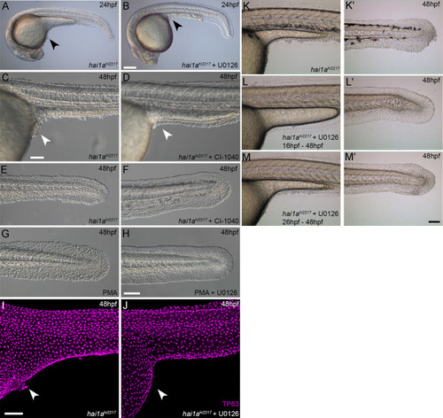
Figure 9—figure supplement 1.

Rescue of the <italic toggle='yes'>hai1a</italic> epidermal phenotype by pERK inhibitors.

(A–F) Lateral DIC images of 24hpf (A, B) or 48hpf (C–F) hai1ahi2217 embryos treated with either DMSO (A, C, E), U0126 (B), or CI-1040 (D, F) showing rescue of general morphology (B), trunk (D), and tail (F) epidermal phenotypes compared to DMSO-treated hai1ahi2217. (G, H) Lateral DIC images of tails of 48hpf WT treated with 125 ng/ml phorbol 12-myristate 13-acetate (PMA) alone (G) or PMA and U0126 (H). (I, J) Lateral projected confocal images of trunks of 48hpf hai1ahi2217 embryos treated with DMSO (I) or U0126 (J) and then fluorescently immunostained for TP63. (K–M′) Lateral DIC images of the trunks (K, L, M) and tails (K′, L′, M′) of 48hpf hai1ahi2217 embryos either untreated (M, M′), treated with 100 µM U0126 from 16hpf to 48hpf (L, L′), or treated with U0126 from 26hpf to 48hpf (M, M′). Whilst treatment from 16hpf led to a complete rescue, initiating treatment later resulted in only partial rescue. Scale bars: (B) = 200 µm; (C, H, I, M′) = 100 µm.

Expression Data

Expression Detail
Antibody Labeling
Phenotype Data

Phenotype Detail
Acknowledgments
This image is the copyrighted work of the attributed author or publisher, and ZFIN has permission only to display this image to its users. Additional permissions should be obtained from the applicable author or publisher of the image. Full text @ Elife