Fig. 1
- ID
- ZDB-FIG-080325-92
- Publication
- Lachnit et al., 2008 - Alterations of the cytoskeleton in all three embryonic lineages contribute to the epiboly defect of Pou5f1/Oct4 deficient MZspg zebrafish embryos
- Other Figures
- All Figure Page
- Back to All Figure Page
|
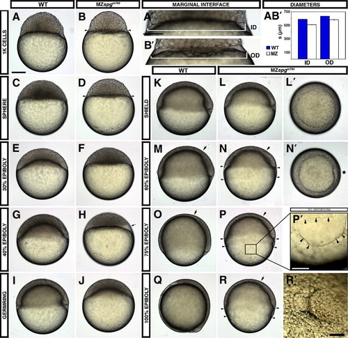
Live phenotype of MZspg embryos reveals severe gastrulation defects. (A?D) At the onset of cell motility, MZspgm793 embryos display a smaller blastoderm with an enhanced constriction between DEL/EVL and YSL and an enlarged YSL (small arrowheads). (A′, B′) Higher magnification of the outer (interface between DEL and YSL) and inner (interface between YSL and YCL) diameter of WT versus MZspgm793. (A, B′) Measurements of the diameters including SE. Inner diameter (ID): MZspgm793: 508 ± 2 μm, WT: 587 ± 4 μm; p = 4 · 10- 10. Outer diameter (OD): MZspgm793 = 576 ± 7 μm, WT = 635 ± 2 μm; p = 1 · 10- 05. (E?H) While the blastoderm in WT starts spreading over the yolk, mutant blastoderm sits on the yolk cell showing translucent deep cell areas (H, arrow). (I, J) A 30% epiboly progress can be detected in MZspgm793 when WT reach germ ring stage. (M, N, N′) At 60% epiboly mutant embryos display a widening gap (arrowheads) between EVL and DEL margin and a small malformed shield (N′, animal view, *). (N, P, R) Vegetally migrating deep cells and anteriorly migrating hypoblast cells in mutant embryos show retardation (arrow marking the leading edge of the anterior mesendoderm). (P2) Higher magnification of the mutant YCL showing cortical clefts (arrowheads); arrows indicate EVL margin. (Q, R) At the end of epiboly, the DEL as well as the EVL appears strongly retarded in MZspgm793. EVL cells seem to have reached about 75?80% epiboly, while deep cells seem not to have passed 50% epiboly. (R2) Animal view of mutant embryos shows a polster like formation. Staging of live embryos has been repeated in three indeE. EVL cells seem to have reached about 75?80% epiboly, while deep cells seem not to have passed 50% epiboly. (R2) Animal view of mutant embryos shows a polster like formation. Staging of live embryos |
| Fish: | |
|---|---|
| Observed In: | |
| Stage Range: | 1k-cell to 90%-epiboly |
Reprinted from Developmental Biology, 315(1), Lachnit, M., Kur, E., and Driever, W., Alterations of the cytoskeleton in all three embryonic lineages contribute to the epiboly defect of Pou5f1/Oct4 deficient MZspg zebrafish embryos, 1-17, Copyright (2008) with permission from Elsevier. Full text @ Dev. Biol.

