
Fig. S5
- ID
- ZDB-FIG-080325-121
- Publication
- Lachnit et al., 2008 - Alterations of the cytoskeleton in all three embryonic lineages contribute to the epiboly defect of Pou5f1/Oct4 deficient MZspg zebrafish embryos
- Other Figures
- All Figure Page
- Back to All Figure Page
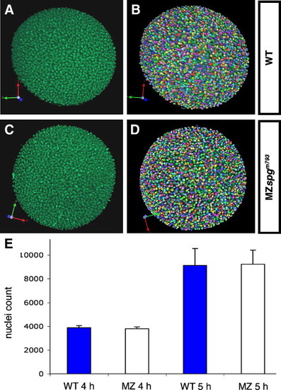
Nuclei counts at same developmental times are similar in wild-type and MZspg mutant embryos during early gastrula stages. Precisely staged embryos were obtained, fixed, and nuclei stained with Sytox Green. (A, C) Z-projections of animal views of wild-type and MZspgm793 mutant embryos. (B, D) Detection of Sytox Green stained nuclei by Volocity software: individual volumes representing stained nuclei are assigned different colors. (E) Nuclei counts for wild-type and MZspg mutant embryos at 4 hpf and 5 hpf. Actual values for individual embryos are: 4 hpf: WT: 4009, 3744, mean: 3877, standard deviation: 187; MZspgm793: 3911, 3683, mean: 3797, standard deviation: 161; 5 hpf: WT: 8154, 8516, 7680, 10804, 10502, mean: 9131, standard deviation: 1424; MZspgm793: 10636, 9220, 8401, 7931, 9984, mean: 9234, standard deviation: 1190. There is no significant difference in nuclei counts between wild-type and MZspgm793mutant embryos. Student's t-test for 5 hpf: t = 0.128, p = 0.9. |
| Fish: | |
|---|---|
| Observed In: | |
| Stage Range: | Sphere to 30%-epiboly |
Reprinted from Developmental Biology, 315(1), Lachnit, M., Kur, E., and Driever, W., Alterations of the cytoskeleton in all three embryonic lineages contribute to the epiboly defect of Pou5f1/Oct4 deficient MZspg zebrafish embryos, 1-17, Copyright (2008) with permission from Elsevier. Full text @ Dev. Biol.