- Title
-
Knockdown of zebrafish YY1a can downregulate the phosphatidylserine (PS) receptor expression, leading to induce the abnormal brain and heart development
- Authors
- Shiu, W.L., Huang, K.R., Hung, J.C., Wu, J.L., Hong, J.R.
- Source
- Full text @ J. Biomed. Sci.
|
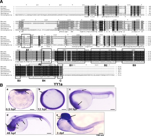
Sequence alignment of zebrafish YY1a and its expression profile. a A sequence alignment of the YY1a protein (where positions of the functional domains are indicated by a series of boxes labeled A-C). Box A contains potential nuclear localization signals (PSGRMKK [118-124 aa] and PRKIKED [227-233 aa]; Fig. 1a, box A1-2). These lie within the predicted intracellular domain (SMART-TMHMM2 program). Box B1-4 contains the potential zinc finger, CH2H2 type, domains (box B1 [CPHKGCTKMFRDNSAMRKHLHTH, residues 241-263, 23 aa], box B2 [CAECGKAFVESSKLKRHQLVH, residues 270-290, 21 aa], box B3 [CTFEGCGKRFSLDFNLRTHVRIH, residues 298-320, 23 aa], and box B4 [CPFDGCNKKFAQSTNLKSHILTH, residues 328-350, 23 aa]), all of which are well characterized as DNA binding modules. Box C (residues 353-356; AKNN for KKXX domain) contains an ER membrane retention signal based on architectural analysis performed with the SMART-TMHMM2 program. b Expression of YY1a from early to late developmental stages detected by in-situ antisense RNA hybridization. YY1a was visualized by blue staining. Lateral view of embryos is shown in panels a-c and e; top view is shown in panel d; anterior is on the right side. (a) One-cell stage (half-hour). YY1a is expressed in all cells examined. b At 12 hpf, YY1a is expressed throughout the embryo. (c) At 24 hpf, YY1a is expressed throughout the embryo, but is especially concentrated within the brain region (indicated by an arrow) and in the anterior part of the embryo. (d) At 48 hpf, YY1a is expressed principally in the posterior notochord (indicated by an arrow) and secondarily in the trunk, brain, kidney, and eyes (indicated by arrows). (e) At 3 dpf, the YY1a is strongly expressed in some major organs (brain, heart, and eyes; indicated by arrows) and mildly expressed throughout the somite. Scale bars denote 100 Ám. EXPRESSION / LABELING:
|
|
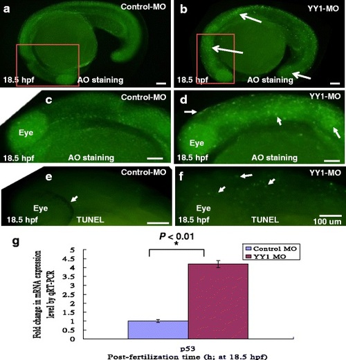
Apoptotic cell analysis by AO staining and TUNEL assay. Control-MO and YY1-MO (25 ng per embryo) were injected at the one-cell stage to block translation of YY1a mRNA. The embryos were fixed and observed at the different stages postfertilization (pf). The AO-stained embryos are shown in a (Control-MO; 18.5 hpf), b (YY1-MO; 18.5 hpf), c (Control-MO; 18.5 hpf; enlarged from A), and d (YY1-MO; 18.5 hpf; enlarged from B; strongly AO-positive cells indicated by arrows). TUNEL stained embryos (all at 18.5 hpf) are shown in e (Control-MO) and f (YY1-MO group). Identification of apoptotic cell death-related gene P53 at 18.5 hpf by qRT-PCR approach as a control is shown in (g). All data were analyzed using either paired or unpaired Student?s t-tests as appropriate. *P < 0.01. The TUNEL-positive cells under the fluorescence microscope are considered apoptotic. Bars indicate 100 Ám EXPRESSION / LABELING:
PHENOTYPE:
|
|
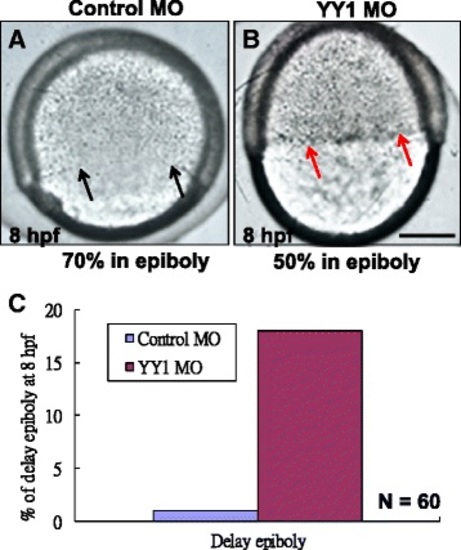
Morpholino-induced knockdown of YY1a prevents cell corpse engulfment and cell migration. a Control-MO-injected embryos, lateral view of a Control-MO-injected embryo about entering into 70 % epiboly stage at 8 hpf (indicated by arrows) reveals the normally developed. b Lateral view of a YY1-MO-injected embryo reveals the delay epiboly about 20 % (indicated by red arrows). Bars indicate 100Ám. c Quantification of the delay epiboly embryos from control-MO and YY1-MO injection groups is shown the delay epiboly migration (N = 60) PHENOTYPE:
|
|
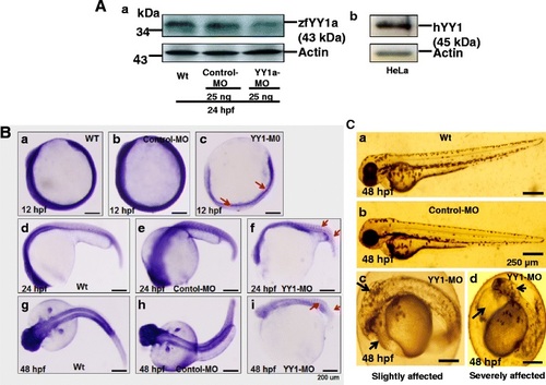
Ubiquitous YY1a inhibition by injection of morpholinos (YY1-MO). a Western-blot analysis of YY1a proteins in the 24-h stage wild type embryos and YY1-MO-injected embryos. The knockdown of YY1a gene in these embryos by YY1-MO injection was shown in panel a, lane 3 (panel a, lanes 1-3; panel b, the positive control, HeLa cell lysate). Actin was the loading control. b Each one-cell stage embryo was injected with either 25 ng of control-MO or 25 ng of YY1-MO. At 12, 24, and 48 hpf, embryos were fixed and examined after in-situ hybridization. The phase-contrast images of the wild type (panels, a, d, and g) and control-MO-injected (panels b, e, and h) embryos show normal development and YY1a expression patterns. However those of the YY1-MO-injected embryos show abnormal development (panels c, f and i) and delayed YY1a expression (panel c, 12 hpf; arrows) and abnormal YY1a expression (panel f and i; 24 and 48 hpf, respectively; arrows). c Phase-contrast images of wild type embryos (panel a), control-MO-injected embryos (panel b), YY1-MO-injected embryos that are slightly affected (panel c), and YY1-MO injected embryos that are severely affected (panel c). The abnormal brain and heart are indicated by arrows. Bars indicate 250 Ám EXPRESSION / LABELING:
PHENOTYPE:
|
|
Morpholino-induced knockdown of YY1a results in down-regulation of PS receptor at 24 hpf. a Western-blot analysis of PSR proteins in lysates of YY1-MO-injected embryos. Note that PSR protein level in these embryos decreases in a YY1-MO dose-dependent manner (Fig. 5a, panel a, lane 1 [control-MO, 50 ng]; lane 2 [YY1-MO, 25 ng], and lane 3 [YY1-MO, 50 ng]). Panel b shows the actin loading control. b One-cell stage embryos were each injected with either 25 ng of control-MO or 25 ng of YY1-MO. At 12, 24, and 48 hpf, embryos were fixed and in-situ hybridization was performed as described in the Methods section. Phase-contrast images show normal development and psr expression patterns in wild type (panels a, d, and g) and control-MO-injected (panels b, e, and h) embryos but abnormal development (panels c, f, and i) and either a mild (panel c, 12 hpf) or severe (panel f, 24 hpf; panel i, 48 hpf) delay in psr expression (arrows) in YY1-MO-injected embryos. Bars indicate 100 Ám EXPRESSION / LABELING:
PHENOTYPE:
|
|
Morpholino-induced knockdown of YY1a results in brain and heart defects. Morphological analysis of embryos injected with either 25 ng of control-MO or YY1-MO and examined at 48 hpf or 3 dpf following staining with pax 2a, nkx 2.5, or no stain. a Panels a-c. The embryos are stained with pax 2a (a, b; top view, anterior to the right) or not stained (d-f; lateral view). The YY1-MO-injected embryos (panel c) has smaller brain (indicated by black arrow) and abnormal pax 2a pattern (indicated by red arrow) as compared with wild type embryos (panel a) and control-MO-injected embryos (panel b). The fore-, mid-, and hindbrains are shorter (indicated by open square [panel f]; cf. with control [panels d and e]). b Staining with nkx 2.5 was used to monitor heart development. Normal heart formation was delayed at 48 hpf (panel c; indicated by star). Compare with the atria (A) and ventricles (V) in panels a and b. The tube-like heart in panel f (indicated by arrows) should be compared with that in wild type and control-MO-injected embryos (panels d and e, respectively) at 3 dpf. Bars indicate 100 Ám EXPRESSION / LABELING:
PHENOTYPE:
|
|
Injection of YY1a mRNA rescued embryos from YY1a morpholino-induced defects. YY1-MO (25 ng) and YY1a mRNA (2.5 ng) were co-injected at the one-to-two cell stage, and embryos were assessed at 48 hpf and 3 dpf. a Panel c shows YY1-MO-injected embryos with severe morphological deformities (indicated by long arrow). After rescue, the deformities were markedly reduced (panel d: indicated by short arrow; cf. wild type [panel a] and control-MO-injected [panel b] embryos). Bars indicate 250 Ám. Whole embryos before (panel g) and after (panel h) rescue can be compared with wild-type (panel e) and control-MO-injected (panel f) embryos. Bars indicate 200 Ám. b The ability of YY1a mRNA injection to rescue embryos from morphological deformities during development at 3 dpf was estimated. c Rescued embryos show normal morphology and normal YY1a expression in the brain, heart, and somites and are all stained with the YY1a probe at 3 dpf developmental stage. The normal morphology and normal YY1a pattern in the brain and heart (indicated by arrows) seen in rescued embryos (panel d) is in contrast to the deformed brains and hearts seen in YY1-MO knockdown embryos (panel c; indicated by arrows). The wild type (panel a) and control-MO-injected (panel b) are the negative controls. Bars indicate 200 Ám EXPRESSION / LABELING:
PHENOTYPE:
|
|
Injection of PSR mRNA rescued embryos from YY1a morpholino-induced defects. YY1-MO (25 ng) and PSR mRNA (1 ng) were co-injected at the one-to-two cell stage, and embryos were assessed at 18.5 hpf, 48 hpf and 3 dpf. a Apoptotic cell analysis by TUNEL assay. Control-MO and YY1-MO (25 ng per embryo) or YY1-MO (25 ng per embryo) plus PSR mRNA (1 ng) were injected at the one-cell stage to express extra PS receptor. The embryos were fixed and observed at the different stages postfertilization (pf). TUNEL stained embryos (all at 18.5 hpf) are shown in A:a (Control-MO), A:b (YY1-MO group) and A:c (extra PSR mRNA group). The TUNEL-positive cells under the fluorescence microscope are considered apoptotic, especially in A:b (indicated by arrows). Bars indicate 100 Ám. (B) Rescued embryos show normal morphology in the brain, heart, and somites. The normal morphology in the brain and heart (indicated by arrows) seen in rescued embryos (panel c, at 24 hpf; f, at 48 hpf; i, at 72 hpf) is in contrast to the deformed brains (indicated by black arrows) and hearts (indicated by red arrows) seen in YY1-MO knockdown embryos (panel b, at 24 hpf; e, at 48 hpf; h, at 72 hpf; indicated by arrows). The control-MO-injected (panel a, at 24 hpf; d, at 48 hpf; g, at 72 hpf) are the negative controls. Bars indicate 200 Ám. c The ability of PSR mRNA injection to rescue embryos from morphological deformities during development at 72 hpf was estimated PHENOTYPE:
|


