- Title
-
Proteomic and functional analysis of NCS-1 binding proteins reveals novel signaling pathways required for inner ear development in zebrafish
- Authors
- Petko, J.A., Kabbani, N., Frey, C., Woll, M., Hickey, K., Craig, M., Canfield, V.A., and Levenson, R.
- Source
- Full text @ BMC Neurosci.
|
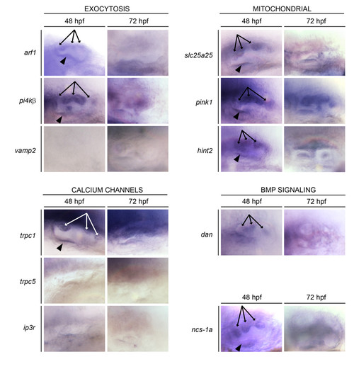
Expression of NCS-1 binding partners (NBPs) during zebrafish inner ear development. Whole mount in situ hybridization analysis was performed at 48 and 72 hpf. Genes are grouped according to presumed functional properties. All images are lateral views of the otic vesicle, anterior to the left. Arrows indicate mRNA expression in semicircular canal protrusions at 48 hpf. Arrowheads indicate mRNA expression in the anterior macula at 48 hpf. EXPRESSION / LABELING:
|
|
GST-pulldowns. GST pulldowns were performed to validate NCS-1/NBP interactions. A human NCS-1-GST fusion protein was used to pull down S-tagged zebrafish NBPs. In each panel, the left (+) lane contains a bacterial lysate expressing the designated S-tagged NBP, the middle lane (PD), contains NBPs pulled down from bacterial lysates after incubation with the NCS-1-GST fusion protein, and the right lane (-) shows proteins pulled down from lysates after incubation with GST alone. Blots were probed with an HRP-conjugated S-tag antibody to detect each S-tagged NBP. Molecular weights (kDa) of S-tagged NPB fragments are indicated to the left of each panel. |
|
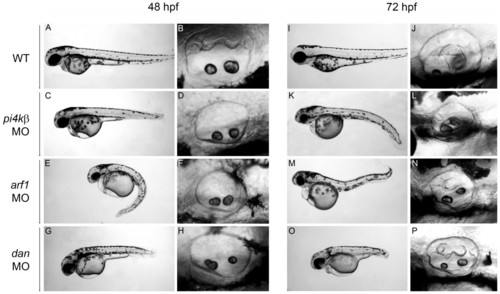
Knockdown of arf-1, pi4kβ, and dan mRNA expression. Antisense MOs targeted against arf-1, pi4kβ, or dan mRNA were injected into single cell embryos, and morphants analyzed at 48 (C-H) and 72 hpf (K-P). For comparison, control (buffer injected) embryos are shown at 48 (A-B) and 72 (I-J) hpf. Embryos were injected with either 4 ng pi4kβ-ATG MO (C-D, K-L), 2 ng arf1-UTR MO (E-F, M-N), or 1ng of dan-ATG MO (G-H, O-P). Images of otic vesicle (B, D, F, H, J, L, N, P) are all lateral views with anterior to the left. Lateral views of wild-type embryos are shown at 48 (A) and 72 (I) hpf. Lateral views of morphant embryos are shown at 48 (C, E, G) and 72 (K, M, O) hpf. PHENOTYPE:
|
|
Phenotypes of embryos co-injected with subeffective doses of NBP MOs. Bar graph depicts percent of fish at 48 hpf or 72 hpf that displayed normal epithelial protrusions or semicircular canal hub formation, abnormal epithelial protrusions or semicircular canal hub formation, or absence of epithelial protrusions. Embryos were injected with 0.5 ng of ncs-1a-ATG MO alone, 0.5 ng of arf1-UTR MO alone, or 2 ng of pi4kβ-ATG MO alone. Alternatively, embryos were co-injected with 0.5 ng of ncs-1a-ATG MO plus 0.5 ng of arf1-UTR MO, 0.5 ng of ncs-1a-ATG MO plus 2 ng of pi4kβ-ATG MO, or 0.5 ng arf1-UTR plus 2 ng pi4kβ-ATG MO. The number of fish assayed for each treatment is displayed above the bars. PHENOTYPE:
|
|
Pink1 expression is required for semicircular canal development. An antisense pink1 MO was used to knock down pink1 mRNA translation. Images are all lateral views of 48 hpf embryos with anterior to the right. (A) head of wild type embryo; (B) otic vesicle of wild type embryo. Head of pink1 morphant injected with 2 ng (C) or 4 ng (E) of pink1-ATG MO. Otic vesicle of pink1 morphant injected with either 2 ng (D) or 4 ng (F) of pink1-ATG MO. PHENOTYPE:
|
|
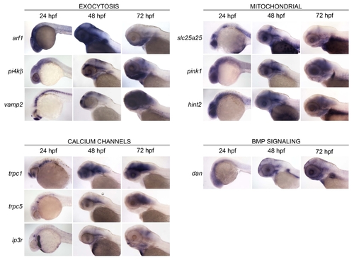
Expression of zebrafish NBPs in head region. Whole mount in situ hybridization analysis was performed at 24, 48, and 72 hpf. Expression profiles in head region are shown for all of the NBPs represented in Figure 1. Genes are grouped according to presumed functional properties. All images are lateral views of the head, anterior to the left. |

Unillustrated author statements EXPRESSION / LABELING:
|


