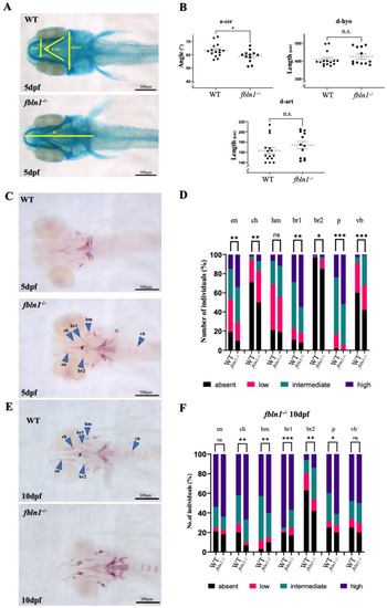
fbln1−/− mutants present increased mineralization at 5 dpf compared to WT. (A) Ventral view of alcian blue stained WT and fbln1−/− larvae at 5 dpf. The distances measured are indicated. (B) fbln1−/− reveal reduced angle between ceratohyals (a-cer) at 5 dpf compared to WT (WT n = 15, fbln1−/− n = 12). (C) Ventral view of alizarin red stained WT and fbln1−/− larvae at 5 dpf. The blue arrowheads point to the skeletal elements: ceratohyal (ch), parasphenoid (p), entopterygoid (en), branchiostegal rays 1 and 2 (br1/br2), hyomandibular (hm), and vertebral body (vb). (D) Fraction (%) of individuals presenting a high (dark blue), intermediate (green), low (red), or absent (black) level of bone mineralization in the different bone elements in WT and fbln1−/− fish at 5 dpf. (WT n = 52, fbln1−/− n = 68). (E) Ventral view of alizarin red stained WT and fbln1−/− larvae at 10 dpf. (F) Fraction (%) of individuals presenting a high (dark blue), intermediate (green), low (red), or absent (black) level of bone mineralization in the different bone elements in WT and fbln1−/− fish at 10 dpf. (WT n = 35, fbln1−/− n = 40). (n.s. = non significant), significance: * p < 0.05, ** p < 0.01, and *** p < 0.001.
|
