
Fig. 3
- ID
- ZDB-FIG-210504-18
- Publication
- Peng et al., 2020 - Induction of Wnt signaling antagonists and p21-activated kinase enhances cardiomyocyte proliferation during zebrafish heart regeneration
- Other Figures
- All Figure Page
- Back to All Figure Page
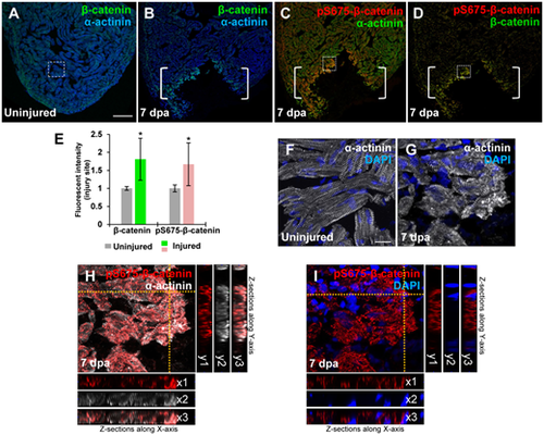
Induction of pS675-?-catenin at disassembled sarcomeres in the injured myocardium following cardiac damage. (A) In uninjured hearts, ?-catenin is detectable throughout the myocardium stained with a sarcomeric Z-disk marker ?-actinin. (B?D) Following ventricular resection, ?-catenin (B) and pS675-?-catenin (C) are induced at the apical cell edge of wounded myocardia. (D) Merged panel of B and C without ?-actinin. Brackets, amputation area. (E) Bar chart depicting ?-catenin and pS675-?-catenin levels following ventricular resection. Fluorescent intensities were measured at the injury border zone using Image J. ?-catenin or pS675-?-catenin levels in control hearts are normalized as 1. Data are mean � SEM from five hearts for each group. Student?s t-test, *P? |