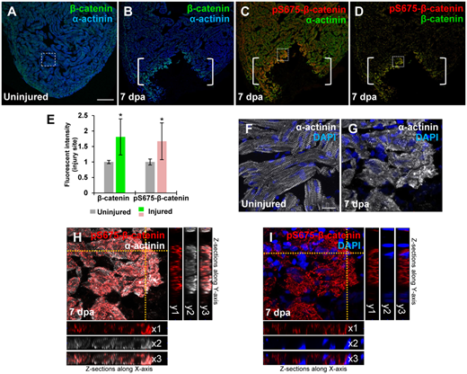
Fig. 3 Induction of pS675-?-catenin at disassembled sarcomeres in the injured myocardium following cardiac damage. (A) In uninjured hearts, ?-catenin is detectable throughout the myocardium stained with a sarcomeric Z-disk marker ?-actinin. (B?D) Following ventricular resection, ?-catenin (B) and pS675-?-catenin (C) are induced at the apical cell edge of wounded myocardia. (D) Merged panel of B and C without ?-actinin. Brackets, amputation area. (E) Bar chart depicting ?-catenin and pS675-?-catenin levels following ventricular resection. Fluorescent intensities were measured at the injury border zone using Image J. ?-catenin or pS675-?-catenin levels in control hearts are normalized as 1. Data are mean ▒ SEM from five hearts for each group. Student?s t-test, *P?
Image
Figure Caption
Acknowledgments
This image is the copyrighted work of the attributed author or publisher, and
ZFIN has permission only to display this image to its users.
Additional permissions should be obtained from the applicable author or publisher of the image.
Full text @ J. Mol. Cell Biol.

