|
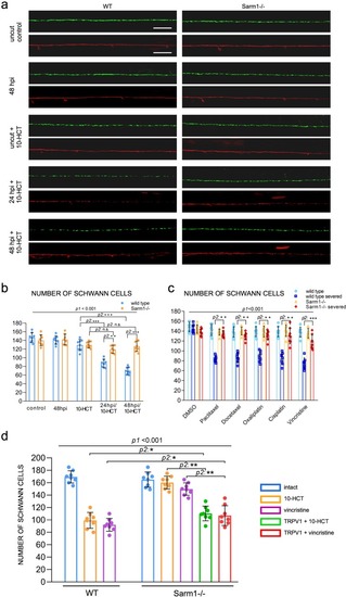
Loss of Sarm1 protects Schwann cells from chemical toxins.a Confocal images showing Schwann cells (green) and lateralis sensory axons (red) in a control specimen (in which axons were not transected), in a specimen 48 h after axon transection, and in specimens treated with 10-HCT (10-hydroxycamptothecin). Left column is wild type and right column shows Sarm1−/−. In all cases, the concentration of 10-HCT in water was 40 μm. Scale bar 100 μm. b Quantification of the Schwann cells from a. Data are shown as mean ± SEM. **p < 0.01, two-way ANOVA, n = 8 (each group), followed by T-test for two individual group. c Quantification of Schwann cells of WT, WT severed, Sarm1−/− and Sarm1−/− severed with the treatment of the indicated chemical compounds for 48 h. Concentrations: Paclitaxel 40 μm, Docetaxel 0.1 μm, Oxaliplatin 500 μm, Cisplatin 50 μm, Vincristine 50 μm. Data are shown as mean ± SEM. *p < 0.05; **p < 0.01, three-way ANOVA, n = 8 (each group), followed by T-test for two individual group. d Quantification of Schwann cells after axon severing, in specimens treated with 10-HCT or Vincristine. The left bar group is wild type. The right bar group is Sarm1−/−, and Sarm1−/− with synthetically eliminated axon segments. Two-way ANOVA, followed by T-test for two individual groups.
|