
Fig. 2
- ID
- ZDB-FIG-190611-28
- Publication
- Turner et al., 2019 - Abrogation of Stem Loop Binding Protein (Slbp) function leads to a failure of cells to transition from proliferation to differentiation, retinal coloboma and midline axon guidance deficits
- Other Figures
- All Figure Page
- Back to All Figure Page
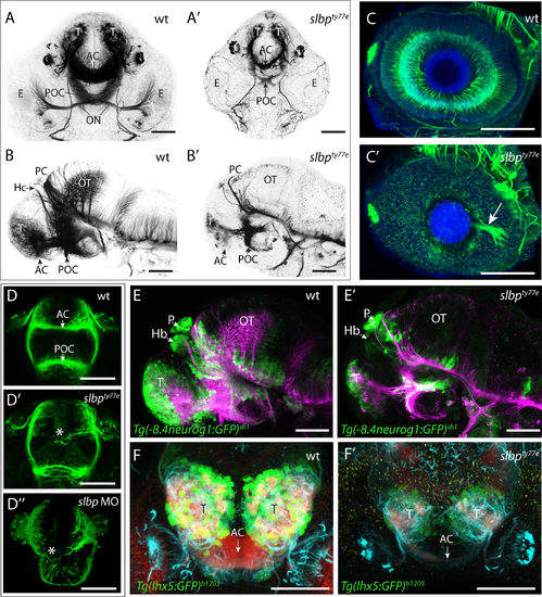
slbpty77e mutants have less neurons and show axonal defects. (A-D”) Acetylated α-tubulin labelling of wildtype (A-D) and ele mutant (A’-D”) embryo brains and eyes. Frontal (A-A’; D-D”) and lateral (B- B’) views of brains and lateral views of eyes (C,C’) of 3dpf (A-C’) and 30hpf (D- D”) wildtype (A-D), slbpty77e mutant (A’-D’) and morphant (D”) embryos. Arrowhead in C’ highlights the aberrant extension of RGC axons within the retina. The asterisks in D’-D” highlight aberrantly positioned axons near the midline commissural region in slbp mutant and morphant embryos. (E-E’) Lateral view with anterior to the left of 3dpf wildtype (E) and slbpty77e mutant (E’) embryos showing expression of the Tg(-8.4neurog1:GFP)sb1 transgene (green) labelled neurons and acetylated α-acetylated tubulin labelled axons/neurites (magenta). (F-F’) Frontal views of the telencephalon in 3dpf wildtype (F) and slbpty77e mutant (F’) embryos showing expression of the Tg(lhx5:GFP)b1205 transgene (green) labelled neurons, SV2 labelled neuropil (red) and acetylated α-acetylated tubulin labelled axons/neurites (cyan). Abbreviations: AC, anterior commissure; POC, post-optic commissure; ON, optic nerve; T, telencephalon; PC, posterior commissure; OT, optic tectum; Hb,habenula; E,eye; P,pineal. Scale bars: (A-C”, E, E’) 100μm (D-D”, F, F’) 50μm |
| Gene: | |
|---|---|
| Antibodies: | |
| Fish: | |
| Knockdown Reagent: | |
| Anatomical Terms: | |
| Stage Range: | Prim-15 to Protruding-mouth |