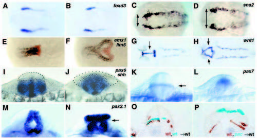
Lack of Ncad causes neurulation defects. (A-N) Whole-mount in situ hybridization for the markers indicated in the top right-hand corner. Dorsal views with anterior towards the left in A-H; optical cross sections dorsal upwards in I-N; all paired panels compare a pac mutant (right) with a wild-type sibling (left). (O,P) Mid-hindbrain cross-sections of wild-type embryos co-transplanted with cells from two different donors, as indicated in the bottom right-hand corner; donor cells are stained in cyan or brown. (A,B) foxd3, three-somite stage; note the identical mediolateral extent of the neural plate in wild-type and pac. (C,D) sna2, five-somite stage; arrows indicate the width of neural plate delineated by sna2 stripes, larger in pac. (E,F) emx1 (blue; marking telencephalon) + lim5 (red; marking posterior diencephalon) (Toyama et al., 1995a), 10- somite stage. In F, cells in the fused part of the lim5 expression domain are located ventrally, cells in the bilateral parts dorsally. (G,H) wnt1, 26 hpf; arrows indicate fused (G) and bilateral (H) expression domains in the midbrain. (I,J) pax6 + shh; 12-somite stage; section at hindbrain level. The alar plate devoid of pax6 staining is outlined by dots. (K,L) pax7, 16-somite stage; optical section at hindbrain level. Arrow in K indicates a pax7 stripe in the interface of basal and alar plate. (M,N) pax2.1, 24 hpf; optical cross-section through midbrain-hindbrain boundary region; arrow indicates the region where basal and alar plate have morphologically separated. (O,P) Chimeric embryos, 24 hpf, cross-section at midbrain (P) and hindbrain (O) levels. Note that in P, wild-type cells (in brown) populate the basal plate, while pacpaR2.10 mutant cells (in cyan) remain alar, although both cell types had initially been transplanted to the same presumptive basal region of the host embryo.
|

