
Fig. 3
- ID
- ZDB-FIG-140717-12
- Publication
- Lele et al., 2002 - parachute/n-cadherin is required for morphogenesis and maintained integrity of the zebrafish neural tube
- Other Figures
- All Figure Page
- Back to All Figure Page
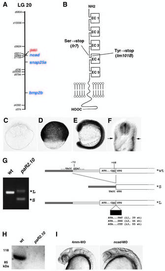
pac encodes N-cadherin. Map positions of the pactm101B mutation and the ncad gene on linkage group 20. Cartoon of the structure of Ncad protein. As indicated, Ncad encoded by pacm101B and pacfr7 displays premature terminations in the extracellular region, shortly after the EC3 domain. (C-F) Expression pattern of ncad. (C) Eight-cell stage, lateral view. (D) 80% epiboly stage, lateral view. (E) Fifteen-somite stage, lateral view. (F) 24 hpf, cross-section at trunk level. ncad expression is restricted to neural tube and slow muscle fibers. (G) RT-PCR analysis of ncad transcripts from pacpaR2.10 mutants, and structure of their 5′ regions. From pacpaR2.10 RNA, no wildtype, but only a predominant smaller transcript (*S) and different larger ncad transcripts (*L) were amplified. (H) Western blot analysis of protein extracts from a pacpaR2.10 and a wild-type sibling embryo with a polyclonal antibody against EC domains 4,5 of zebrafish Ncad (Bitzur et al., 1994). The wild-type extract gives an Ncad-positive band of the expected size, whereas no such band is seen in extracts from the pacpaR2.10 mutant. Before antibody incubation, the blot had been stained with Ponceau Red, showing that equal amounts of proteins were loaded in both lanes. Similar analyses failed to detect Ncad protein from pacfr7 mutants, as expected, because, according to the cDNA sequence, pacfr7 mutant protein terminates after the third EC domain. (I) Phenocopy of the pac mutant phenotype with anti-ncad morpholino ncad-MO1 (right, compare with Fig. 1F) and with the four-mismatch control MO (left) (see Materials and Methods). Embryos at 24 hpf, lateral view of head region. |
| Fish: | |
|---|---|
| Knockdown Reagent: | |
| Observed In: | |
| Stage: | Prim-5 |