
Fig. 4
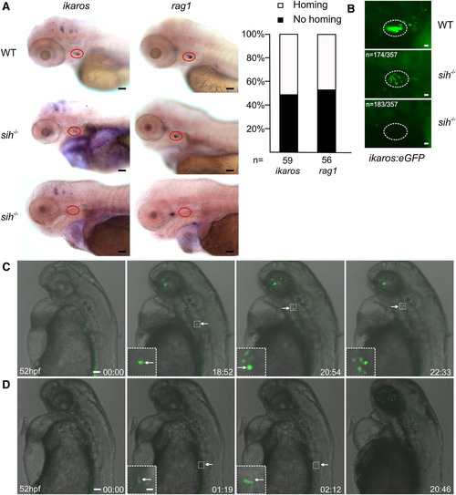
Blood Circulation Is Not Required for Thymus Homing (A) Whole-mount RNA in situ hybridization with antisense ikaros- and rag1-specific probes of wild-type (wt; top row) and silent heart mutant (sih-/-; middle and bottom rows) embryos at 4 dpf. The thymus is highlighted by red circles; lateral views. About half of sih-/- embryos contained at least one positive cell in the thymic rudiment at this time point (histograms at right). Representative results for 70 wild-type and 59 and 56 mutant embryos. Scale bars represent 50 μm. (B) A similar analysis of WT and sih-/- fish additionally transgenic for ikaros:eGFP yielded similar results. Representative results in <400 wild-type and 174 and 183 mutant embryos. Scale bars represent 10 μm. (C) Still photographs from a time-lapse recording tracking a thymus-settling precursor in a sih-/- mutant with eventual thymus colonization. The still series begins at 52 hpf (00 hr:00 min); several other time points are shown including high-power views of relevant regions (insets). Representative results for four embryos. Scale bars represent 50 μm for overviews, 10 μm for insets. See also Movie S5. (D) Still photographs from a time-lapse recording of a sih-/- mutant without thymus colonization. The cell shown at 01:19 and 02:12 time points is a myeloid cell, which shows normal migratory behavior. Representative results for four embryos. Scale bars represent 50 μm for overviews, 10 μm for insets. See also Movie S6. |
| Genes: | |
|---|---|
| Fish: | |
| Anatomical Terms: | |
| Stage Range: | Long-pec to Day 4 |
| Fish: | |
|---|---|
| Observed In: | |
| Stage Range: | Long-pec to Day 4 |
Reprinted from Immunity, 36(2), Hess, I., and Boehm, T., Intravital Imaging of Thymopoiesis Reveals Dynamic Lympho-Epithelial Interactions, 298-309, Copyright (2012) with permission from Elsevier. Full text @ Immunity