
Fig. S6
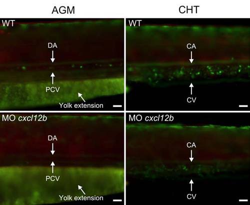
Reduced numbers of ikaros-expressing cells in hematopoietic tissues in cxcl12b morphants �Embryos double-transgenic for ikaros:eGFP and foxn1:mCherry were injected with a cxcl12b-specific anti-sense morpholino and assayed at 63 hpf for green cells in the AGM (aorta-gonad-mesonephros and CHT (caudal hematopoietic tissue) regions. Photographs representative of ~250 wild-type and 48 mutant embryos. Quantitative assessment (values shown are mean � s.e.m.) revealed significant (Fisher�s exact probability test) reductions in both types of tissues. AGM: wild-type fish exhibit 8�0.58 (n=3) and cxcl12b morphants exhibit 0.75�0.25 (n=4) ikaros+ cells per high-power field (P<0.001); CHT: wild-type fish exhibit 32�3.5 (n=3) and cxcl12b morphants exhibit 8.5�1.3 (n=6) ikaros+ cells per high-power field (P<0.001). Anatomical landmarks are indicated: PCV, posterior cardinal vein; CV, caudal vein; CA, caudal artery; DA, dorsal aorta. Scale bars, 50μm. |
| Fish: | |
|---|---|
| Knockdown Reagent: | |
| Observed In: | |
| Stage: | Pec-fin |
Reprinted from Immunity, 36(2), Hess, I., and Boehm, T., Intravital Imaging of Thymopoiesis Reveals Dynamic Lympho-Epithelial Interactions, 298-309, Copyright (2012) with permission from Elsevier. Full text @ Immunity