FIGURE SUMMARY
Title

Selective Histone Deacetylase 6 Inhibitors Restore Cone Photoreceptor Vision or Outer Segment Morphology in Zebrafish and Mouse Models of Retinal Blindness

Authors
Sundaramurthi, H., Roche, S.L., Grice, G.L., Moran, A., Dillion, E.T., Campiani, G., Nathan, J.A., Kennedy, B.N.
Source
Full text @ Front Cell Dev Biol

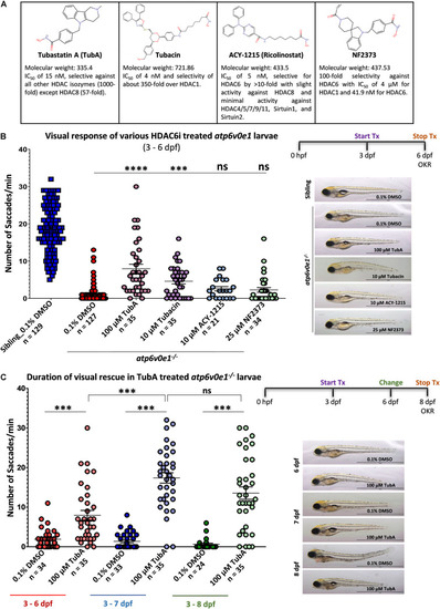
Selective HDAC6i significantly restores visual function in atp6v0e1–/–, a zebrafish model of retinal blindness. (A) Table describing the physiochemical properties of the four HDAC6i candidates selected for present study. Chemical structures were drawn using Chemspider (http://www.chemspider. com/StructureSearch.aspx). (B)atp6v0e1–/– mutants were treated at maximum tolerated concentration (refer to Supplementary Figures S1, S2A) of each HDAC6i, from 3–6 days post fertilization (dpf). Visual function was markedly improved upon treatment with 100 μM TubA, 10 μM Tubacin, 10 μM ACY-1215 or 25 μM NF2373 compared to vehicle-control (0.1% DMSO) treated larvae. Panel on right shows representative larval wholemount images. Statistical analysis was performed using One-way ANOVA with Dunnett’s multiple comparisons, where ∗∗∗p-value = 0.0008 and **** means a p-value of ≤ 0.0001. N = 3 and n = 12 per treatment group. (C) The duration of action of TubA was determined, whereby atp6v0e1–/– larvae were treated from 3 to 8 dpf, and visual function measured at 6, 7 or 8 dpf (refer to Supplementary Figure S2B for visual response in siblings). A significant increase in visual response was recorded at 7 and 8 dpf, respectively. Representative larval images presented in the right panel. Student’s T test was used for statistical analysis, where ∗∗∗ means a p-value of ≤ 0.0001. N = 3 and n = 12 per treatment group.

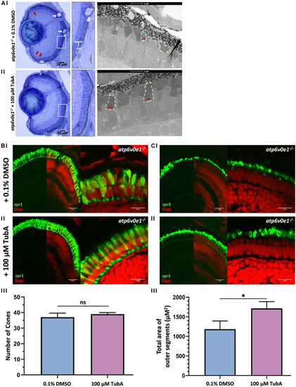
TubA treatment improved retinal morphology in atp6v0e1–/– larvae. (Ai) LM images revealed large vacuoles (pink arrows) in the RPE layer, pyknotic nuclei (red arrows) in the CMZ and shortened photoreceptor outer segments in vehicle-control treated atp6v0e1–/– mutants. (Aii) The photoreceptor outer segments were observed to be more elongated, more prominent, and better organized in TubA-treated atp6v0e1–/– mutants compared to vehicle-control treated atp6v0e1–/– mutants (Supplementary Figure S3). (B,C) Confocal images of immuno-stained vehicle-control or TubA-treated atp6v0e1–/– mutant retina with cone (zpr-1) photoreceptor and rod and cone outer segment (zpr-3) specific markers. (Bi,Ci) Panels present aberrant cone and rod outer segment morphology in vehicle-control treated atp6v0e1–/– mutants. (Bii,Cii) TubA treatment improved photoreceptor structural integrity and organization in atp6v0e1–/– mutants. (Biii,Ciii) Graphical presentation of the number of cone cells (zpr-1 staining) and total area occupied by photoreceptor outer segments (zpr-3 staining), respectively (refer to Supplementary Figure S4 for analysis of retinal histology in siblings; 3 sections per sample was analyzed, N = 3). Student’s unpaired T-test was used for statistical analysis.

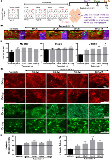
TubA preserves cone cells in rd10 mice retinal explants. (A) Schematic diagram outlining the drug treatment protocol in retinal explants from homozygous rd10/rd10 mice (B6.CXBI–Pde6brd10/J) at postnatal day 14 (P14). (B) Retinal section fluorescent images of explants treated with increasing concentrations of TubA or vehicle control, labeled for rhodopsin (rods) and cone arrestin (cones). The total number of nuclei in the outer nuclear layer (ONL) did not change significantly across the TubA treatment group. Likewise, the number of rhodopsin positive outer segments did not significantly change. A dose-dependent increase in the number of cone arrestin positive outer segments was observed, with a significant increase following 100 μM TubA treatment. (C) In wholemount preparations, no significant increase in fluorescence intensity of rhodopsin positive cells, was observed following TubA treatment. A significant increase in the number of cells with cone arrestin positive outer segments was identified at 50 μM and 100 μM TubA treatment. Statistical analysis was performed using One-way ANOVA with Dunnett’s multiple comparisons. N = 4 per treatment group.

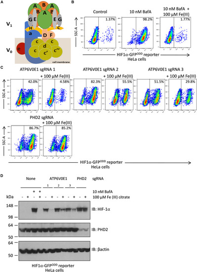
Iron supplementation restores HIF-1α levels to normal following ATP6V0E1 inhibition in HeLa cells. (A) Schematic diagram of the multimeric V-ATPase complex. (B) Chemical inhibition of V-ATPase by 10 nM BafA treatment for 24 h, increased HIF-1α levels in HIFα-GFPODD reporter cells. Treatment with 100 μM Fe (III) citrate significantly reduced the elevated HIF-1α levels associated with loss of ATP6V0E1 (1 × 10⁶ cells per sample harvested and analyzed; N = 2). (C) Knock-down of ATP6V0E1 subunit with three different CRISPR-Cas9 guides resulted in significant upregulation of HIF-1α levels in HIFα-GFPODD reporter cells. Co-treating cells with 100 μM Fe (III) citrate led to a reduction in HIF-1α levels across the three ATP6V0E1 depleted cells. phd2 was knocked down as a control and treatment with 100 μM Fe (III) citrate did not result in reduction of HIF-1α levels (1 × 10⁶ cells per sample harvested and analyzed; N = 2). FACs plot shown is a representative image of two biological repeats performed. (D) Immunoblot analysis for HIF-1α and PHD2 levels in HIFα-GFPODD reporter cells with either ATP6V0E1 or PHD2 depleted or treated with 10 nM BafA. The cells were treated with 100 μM Fe (III) citrate for 24 h. β actin was used as a control. Results validated findings observed by flow cytometry, whereby HIF-1α levels were upregulated following ATP6V0E1 knock-down or inhibition and levels were re-normalized upon Fe (III) citrate treatment. Treatment of Fe (III) citrate in PHD2 depleted cells did not alter HIF-1α levels. All experiments were performed in biological duplicate.

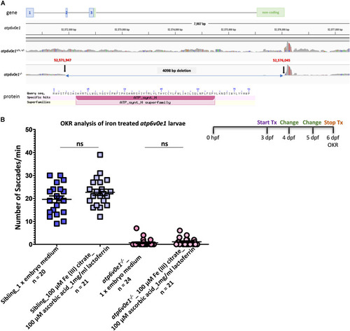
Iron supplementation did not restore vision in atp6v0e1 mutants. (A) Schematic diagram depicting the 4,098 bp deletion in the atp6v0e1 subunit identified through whole genome sequencing. (B) Treatment of atp6v0e1–/– with 100 μM Fe (III) citrate, co-supplemented with 100 μM ascorbic acid and 1 mg/ml of lactoferrin daily for 3 days, did not restore visual function as measured by OKR. Experiments were performed in duplicate and Student’s T-Test was used for statistical analysis.

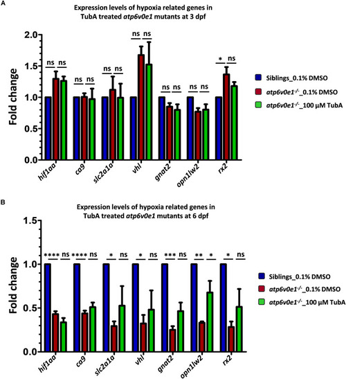
Analysis of hif1aa and targets expression levels in atp6v0e1–/– treated with TubA. (A)hif1aa and select hif1aa downstream target gene(s) expression levels were quantified by qRT-PCR at 4 hpt (i.e., at 3 dpf). No significant changes in hif1aa, slc2a1a, vhl or ca9 transcript levels occurred in atp6v0e1–/– in comparison to vehicle-control treated siblings. Treatment with TubA did not significantly alter these transcript levels either. In atp6v0e1–/–, retinal-specific genes gnat2 and opn1lw2 were not significantly changed but rx2 was significantly upregulated. Upon treatment with TubA, no significant reduction in rx2 transcript levels was detected. (B) Similarly, hif1aa and select hif1aa downstream target gene(s) expression levels were quantified at 3 dpt (i.e., at 6 dpf). A significant reduction in hif1aa, ca9, slc2a1a and vhl transcript levels was identified in vehicle-control treated atp6v0e1–/–. Retinal-specific genes gnat2, opn1lw2 and rx2 were also significantly downregulated compared to vehicle-control treated siblings. TubA-treatment of atp6v0e1–/– resulted in no significant change in hif1aa, ca9, slc2a1a and vhl levels. opn1lw2 transcript levels were significantly increased following TubA-treatment. All experiments were performed in triplicates (n = 60 eyes/treatment condition) and one-way ANOVA with Dunnett’s multiple comparisons was used for statistical analysis.

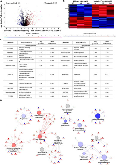
Multiple pathways are implicated in the disease pathomechanism of atp6v0e1–/– as identified by proteome profiling. (A,B) Volcano plot and heatmap representing 790 differentially expressed proteins between vehicle-control treated siblings and atp6v0e1–/–, out of which, 119 proteins were upregulated (in red) and 50 downregulated (in blue) significantly, given a cut-off of fold change ± 1.2 and log p-value ≥ 1.3. Student’s T-Test was used for statistical analysis. (C) Table describing the top-most significantly downregulated and upregulated proteins. (D) KEGG pathway analysis of significantly downregulated and upregulated proteins in vehicle-control treated atp6v0e1–/– compared to vehicle-control treated siblings.

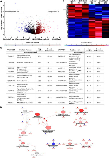
TubA plays a multifaceted role to improve visual function and preserve photoreceptors. (A,B) A total of 347 proteins were identified to be differentially expressed between 3–6 dpf vehicle-control and TubA-treated atp6v0e1–/–. 23 proteins were upregulated (red) and 50 proteins downregulated (blue), with a cut-off of fold change ± 1.2 and –log p-value ≥ 1.3, as shown by the volcano plot and heatmap. Student’s T-Test was used for statistical analysis. (C) Table listing the most significantly down- and upregulated proteins. (D) Graphical representation of significantly downregulated and upregulated pathways in TubA-treated atp6v0e1–/– as identified by KEGG pathway analysis.

Acknowledgments
This image is the copyrighted work of the attributed author or publisher, and ZFIN has permission only to display this image to its users. Additional permissions should be obtained from the applicable author or publisher of the image. Full text @ Front Cell Dev Biol