FIGURE SUMMARY
Title

Impaired brain homeostasis and neurogenesis in diet-induced overweight zebrafish: a preventive role from A. borbonica extract

Authors
Ghaddar, B., Veeren, B., Rondeau, P., Bringart, M., Lefebvre d'Hellencourt, C., Meilhac, O., Bascands, J.L., Diotel, N.
Source
Full text @ Sci. Rep.

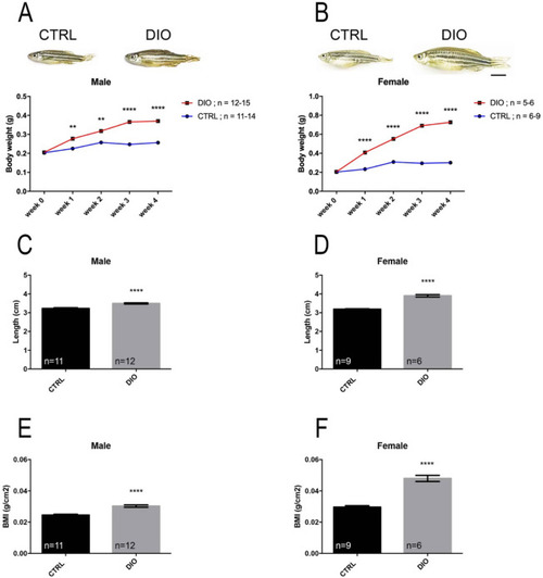
DIO results in increased body weight, length and BMI in both male and female zebrafish. (A), (B) Graphs illustrating the body weight measurements during 4 weeks for both CTRL and DIO-treated zebrafish in male and female, respectively. The zebrafish pictures highlight the morphological differences at week 4. (C), (D) Body length measurements at week 4 in male and female zebrafish, respectively. (E), (F) Body mass index (BMI; grams per square centimeter) calculated at week 4. n = number of fish. One-way ANOVA (A, B) and Student's t-test (CF): **p < 0.01; ****p < 0.0001. Error bars correspond to standard error of the mean (SEM). Scale bar: 7 mm.

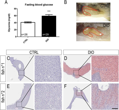
DIO leads to increased fasting blood glucose levels and to liver steatosis in zebrafish. (A) Fasting blood glucose measurements at week 4 in CTRL and DIO-treated fish. (B) Zebrafish liver pictures highlighting the increased size and yellowish color of liver in DIO-treated fish compared to CTRL. (C), (E) Liver sections stained with Oil Red O in two control fish showing no lipid accumulation. (D), (F) Liver sections stained with Oil Red O in two DIO-treated fish showing different levels of lipid accumulation (red color) and highlighting liver steatosis. Nuclei were stained with hematoxylin (CF). These pictures are representative of 3 fish studied. n = number of fish. Student's t-test: ***p < 0.001. Error bars correspond to standard error of the mean (SEM). Scale bar: 1.5 mm (A); 250 μm for lower magnification pictures, 50 μm for higher magnification ones (CF).

DIO increases BBB leakage and neuroinflammation. (A) Dorsal view pictures of the CTRL and DIO zebrafish brains following Evans blue staining. Note the blue staining observed in DIO-treated fish compared to controls (n = 6 brains). (BE) qPCR gene expression analysis of nfkb and pro-inflammatory cytokines (il1β, il6 and tnfα) in CTRL and DIO fish (n = 3 pools of 2 brains). (F) Couting of ramified and amoeboid microglia in the ventral telencephalon (Vv Vd) and the anterior hypothalamus (Hv) regions in both CTRL and DIO-treated fish. (G) Representative L-plastin immunohistochemistry pictures showing microglia morphology in CTRL and DIO zebrafish brain in the subpallium (Vv Vd). Note the apparent increase in amoeboid-like morphology and the stronger L-plastin staining intensity in DIO fish compared to the control ones. n = number of fish. Student's t-test: * p < 0.05; **p < 0.01; ***p < 0.001. Error bar: standard error of the mean (SEM). Scale bar: 0.8 mm (A); 148 μm (G).

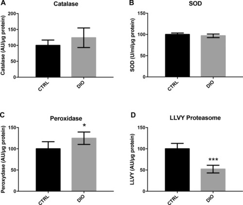
DIO disrupts antioxidant enzymes and proteasome activity in the brain of adult zebrafish. (AD) Cerebral catalase, total superoxide dismutase (SOD), peroxidase and LLVY proteasome activities performed in lysates of adult zebrafish brain. n = 7 from two independent experiments. Student's t-test: *p < 0.05 ***p < 0.001. Error bar: standard error of the mean (SEM).

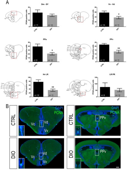
DIO impairs neurogenesis in the forebrain of adult zebrafish. (A) Statistical analysis of the number of proliferative cell (PCNA-positive) in CTRL and DIO-treated zebrafish. The respective brain schemes correspond to the transversal sections of the zebrafish brain for each studied region showing the main brain domains/nuclei according to the Zebrafish Brain Atlas from Wullimann et al. and were adapted from Menuet et al.84,85. A significant decrease in proliferative activity was observed between CTRL and DIO zebrafish in the Vv Vd, PPa, PPv and Hv LR neurogenic regions. (B) Representative digital pictures of PCNA immunohistochemistry (green) and cell nuclei counterstaining (DAPI in blue) on cryostat brain sections of CTRL (up) and DIO-treated fish (down). n = number of brains studied pooled from two independent experiments. Student's t-test: *p < 0.05 **p < 0.01. Error bar: standard error of the mean (SEM). Scale bar = 32 μm. Vv: ventral nucleus of ventral telencephalic area; Vd: dorsal nucleus of ventral telencephalic area; Dm: medial zone of dorsal telencephalic area; PPa: parvocellular preoptic nucleus, anterior part; PPv: periventricular pretectal nucleus; Hv: ventral zone of periventricular hypothalamus; LR: lateral recess of diencephalic nucleus; PR: posterior recess of diencephalic ventricle.

DIO increases the occurrence of inactivity in zebrafish. (A) graph showing the increased number of inactivity state (locomotion < 4 mm/sec) in DIO fish compared to controls, normalized to 100%. (B) graph showing that the total distance traveled during 10 min is similar between CTRL and DIO fish. n = 12 from 3 independent experiments. Student's t-test: *p < 0.05. Error bar: standard error of the mean (SEM).

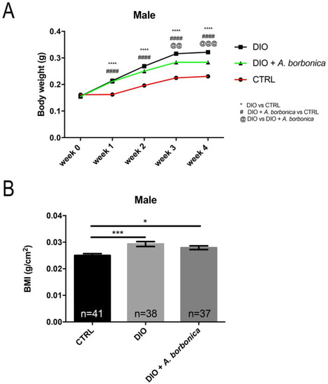
A. borbonica aqueous extracts slightly prevents from body weight gain and excessive BMI increase induced by DIO. (A) Line graph showing the increase in body weight of CTRL, DIO and DIO + A. borbonica during 4 weeks. (B) Graph showing the BMI of all groups at week 4. n = number of fish. One-way ANOVA (A) and Student's t-test (B): *p < 0.05 ***p < 0.001. Bar graph: standard error of the mean (SEM).

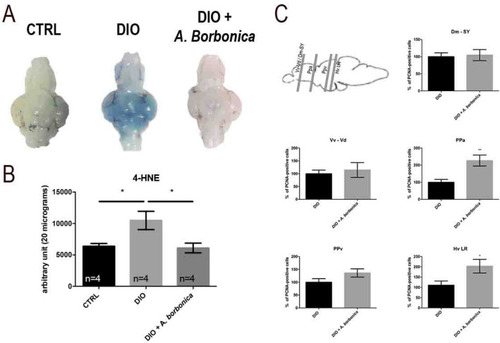
A. borbonica aqueous extracts treatment prevents from BBB leakage, brain oxidative stress and partially protects from the decreased neurogenesis induced by DIO. (A) Dorsal views of zebrafish brains after Evans Blue dye injection in CTRL, DIO, and DIO + A. borbonica treated fish. Zebrafish brains remain mostly white except for the DIO brains. (B) Dot blot quantification showing that the A. borbonica treatment in DIO fish prevents the cerebral increase in 4-HNE levels induced by overfeeding. n = number of brain fish studied. One-way ANOVA: *p < 0.05. Bar graph: standard error of the mean (SEM). (C) Statistical analysis of the number of proliferative cell (PCNA-positive) in DIO and DIO + A. borbonica treated zebrafish normalized to 100%. The sagittal brain scheme shows the corresponding section through the Dm-SY/Vv-Vd, the PPa, the PPv and the Hv LR. A weak significant increase in proliferative activity was observed between DIO and DIO + A. borbonica in the preoptic area (PPa) and the hypothalamic region (Hv LR). n = 8–11 fish. Student's t-test: *p < 0.05 **p < 0.01. Bar graph: standard error of the mean (SEM).

Acknowledgments
This image is the copyrighted work of the attributed author or publisher, and ZFIN has permission only to display this image to its users. Additional permissions should be obtained from the applicable author or publisher of the image. Full text @ Sci. Rep.