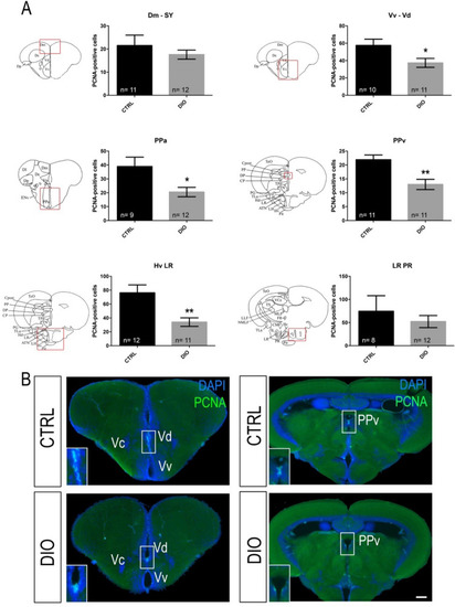
DIO impairs neurogenesis in the forebrain of adult zebrafish. (A) Statistical analysis of the number of proliferative cell (PCNA-positive) in CTRL and DIO-treated zebrafish. The respective brain schemes correspond to the transversal sections of the zebrafish brain for each studied region showing the main brain domains/nuclei according to the Zebrafish Brain Atlas from Wullimann et al. and were adapted from Menuet et al.84,85. A significant decrease in proliferative activity was observed between CTRL and DIO zebrafish in the Vv Vd, PPa, PPv and Hv LR neurogenic regions. (B) Representative digital pictures of PCNA immunohistochemistry (green) and cell nuclei counterstaining (DAPI in blue) on cryostat brain sections of CTRL (up) and DIO-treated fish (down). n = number of brains studied pooled from two independent experiments. Student's t-test: *p < 0.05 **p < 0.01. Error bar: standard error of the mean (SEM). Scale bar = 32 μm. Vv: ventral nucleus of ventral telencephalic area; Vd: dorsal nucleus of ventral telencephalic area; Dm: medial zone of dorsal telencephalic area; PPa: parvocellular preoptic nucleus, anterior part; PPv: periventricular pretectal nucleus; Hv: ventral zone of periventricular hypothalamus; LR: lateral recess of diencephalic nucleus; PR: posterior recess of diencephalic ventricle.
|
