- Title
-
The ciliopathy protein TALPID3/KIAA0586 acts upstream of Rab8 activation in zebrafish photoreceptor outer segment formation and maintenance
- Authors
- Naharros, I.O., Cristian, F.B., Zang, J., Gesemann, M., Ingham, P.W., Neuhauss, S.C.F., Bachmann-Gagescu, R.
- Source
- Full text @ Sci. Rep.
|
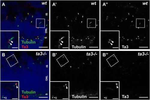
Ta3 localizes to mother and daughter centrioles of photoreceptor primary cilia and is mostly absent in zygotic ta3 mutant PRs. (A?A?) 3 dpf retinal cryosections stained with anti-acetylated tubulin to mark the nascent cilium (green) and with anti-Ta3 (red) show localization of Talpid3 at the base of PR cilia in wildtype (wt) larvae. The double arrowheads highlight Ta3 localization at mother and daughter centriole. (B?B?) In 3 dpf cryosections from ta3?/? larvae, the Ta3 signal is mostly abolished (empty arrowhead in (B?)), but isolated BBs still have positive Ta3 signal, likely maternally deposited (arrowhead in (B?)). The boxed areas in (A?B?) are shown as an inset at the bottom left of each corresponding panel. ONL outer nuclear layer, OS outer segment, S synapse. Scale bars: 2.5?Ám in (A?B?), 1?Ám in insets. EXPRESSION / LABELING:
PHENOTYPE:
|
|
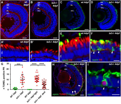
ta3 mutants show progressive retinal degeneration and intracellular opsin accumulation. (A,B) Retinal lamination is unaffected in ta3 mutants as seen on cryosections at 4 dpf stained with the lipophilic dye BODIPY (red) to mark cell membranes and outer segments and DAPI (blue) to mark nuclei. Note the reduced number of PRs with OSs and the cell shape changes in ta3 mutants compared to wildtype (wt) (A??B?). (C,D) Marked intracellular opsin accumulation on 4 dpf cryosections stained with 4D2 antibody (green) recognizing rhodopsin and red-green cone opsin on whole eye cryosections. Nuclei are counterstained with DAPI. (E,F) High magnification images of the PR cell layer of wt (E) and ta3 mutant (F) cryosections at 4 dpf stained with 4D2 antibody (green in E,F) and with BODIPY (red in E,F) to highlight the OSs. Note the substantial intracellular opsin accumulation in mutant PRs in (F) (arrowheads). (G) Progressive PR cell death in ta3 mutants as assessed with a TUNEL assay. Note the significantly smaller amount of TUNEL positive cells in 4 dpf ta3?/? larvae raised in darkness (dark red inverted triangles) compared to those raised in a normal light cycle (light red triangles). Quantification was performed on confocal stacks of identical thickness from cryosections of whole retinas through equivalent regions of the eyes. Each datapoint indicates the number of TUNEL positive nuclei counted in a single larva. ***p?<?0.001, ****p?<?0.0001, Student?s t-test. Bars represent standard deviation. (H) Whole ta3?/? eye cryosection stained with TUNEL assay (red) and with 4D2 antibody (green). Arrowheads mark TUNEL positive PRs. (I) Higher magnification image of a PR with substantial opsin mislocalization (4D2, green) and positive TUNEL reaction (red, arrowhead). dpf days post fertilization, GCL granule cell layer, INL inner nuclear layer, IPL inner plexiform layer, n number, NS not significant, ONL outer nuclear layer, OPL outer plexiform layer, OS outer segment, PR photoreceptor, S synapse, wt wild-type. Scale bars: 30?Ám in (A,B) and H, 4?Ám in (A??B?), (E,F) and I, 40?Ám in (C,D). EXPRESSION / LABELING:
PHENOTYPE:
|
|
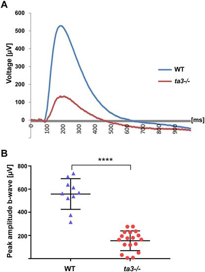
Loss of Ta3 leads to visual function loss. (A) Representative electroretinogram (ERG) recording of wildtype (WT, blue curve) and ta3 mutant (red curve) 6 dpf larvae exposed to light stimuli of 7000 lux (bright light intensity). (B) Average b-wave peak responses for WT (blue triangles) and ta3 mutants (red circles) (n?=?18 ta3 mutant animals and 10 WT animals). Bars represent standard deviation. ****p? PHENOTYPE:
|
|
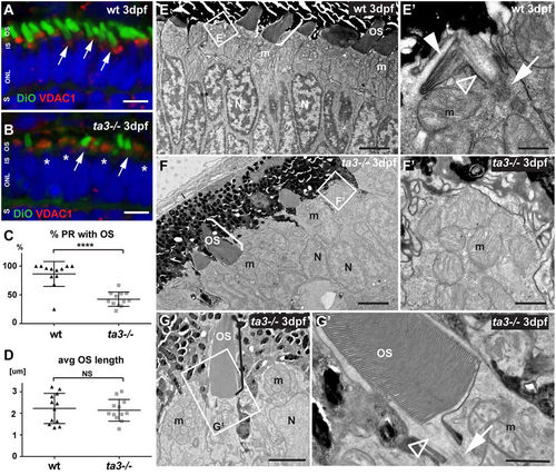
Deficient OS development in ta3?/? larvae in the majority of PRs. (A,B) 3 dpf cryosections stained with DiO to mark the OS (green, arrows) and with VDAC1 to mark mitochondrial clusters show reduced numbers of OSs in ta3 mutants (B) compared to wildtype (wt) (A). Asterisks highlight PRs without OS in mutants. Nuclei are counterstained with DAPI (blue). (C) Quantification of the proportion of PRs with extended OSs in wt (black triangles) compared to ta3 mutants (grey squares) shows significantly reduced PRs with OSs in mutants (****p?<?0.0001, t-test, n?=?12 wt and 12 mutant larvae). Each data point represents the proportion of PRs with OSs on a single confocal section of a single larva. Bars are standard deviation. (D) Quantification of average OS length on cryosections shows no significant difference between 3 dpf wt and ta3 mutants (p?=?0.7366, Student?s t-test, n?=?12 wt and 12 mutant larvae). Each data point represents the average OS length on a single confocal section of a single larva. Error bars are standard deviation. (E?G?) Representative transmission electron microscopy images of 3 dpf wt (E?E?) and ta3 mutant (F?G?) retinae. (E?E?) The majority of wt PRs have extended OSs (bracket in E) or are starting to stack membranes to do so (white arrowhead in E?). Note the BB (arrow in E?) and the connecting cilium (empty arrowhead in E?) in wt photoreceptors. (F?F?) In contrast, in ta3?/? PRs, only a minority of PRs have extended OSs (bracket in F), and no attempt at membrane stacking is observed in the other PRs (F?). (G) Note the normal appearance of the OSs that have extended in the mutants, including the structurally normal connecting cilium (arrowhead in G?) and basal body (arrow in G?). avg average, dpf days post fertilization, IS inner segment, m mitochondria, N nuclei, NS not significant, ONL outer nuclear layer, OS outer segment, PR photoreceptor, S synapse, wt wild-type. Scale bars: 4?Ám in (A,B), 3?Ám in E and F, 0.5?Ám in (E?), 2?Ám in (G), 1?Ám in (F??G?). EXPRESSION / LABELING:
PHENOTYPE:
|
|
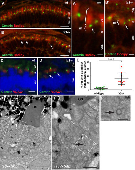
Abnormal BB localization in ta3?/? PR underlies the OS development defect. (A,B) 4 dpf cryosections stained with BODIPY (red) marking membranes of the OS and mitochondrial cluster and with anti-Centrin antibody (green) to mark the basal bodies, show aberrant localization of BB below the mitochondria in ta3?/? PRs. In wildtype (wt) PRs, the centrin-marked BB (arrow in A?) is located just basal to the OS (straight bracket) and apical to the mitochondrial cluster (curved bracket), while this localization is lost in a substantial number of ta3 mutant PRs with BBs located below the mitochondrial cluster (arrow in B?). (C,D) Immunofluorescence on 4 dpf cryosections with anti-VDAC1 antibody (red) to mark the mitochondria and anti-Centrin antibody to mark the BB showing aberrant positioning of BBs in ta3 mutants basal to the mitochondria (arrows in D) compared to wt (C). (E) Quantification of BB position with respect to the mitochondrial cluster based on immunofluorescence with anti-Centrin antibody. Each data point represents the proportion of PRs with BBs below the lower 1/3 of the mitochondrial cluster on one confocal section of a whole retina from one single larva. The proportion of 4 dpf PRs with aberrant BB positioning below the mitochondrial cluster is significantly increased in ta3 mutants (****p?<?0.0001, Student?s t-test, n?=?10 wildtype and 9 mutant larvae). Bars are standard deviation. (F?F?) Representative TEM images showing presence of a BB in cross-section (arrow) within a mitochondrial cluster even at the later stage of 5 dpf, when all BBs should have docked to the apical membrane. (F?) Is the boxed area in (F) and (F?) is the boxed area in (F?). BB basal body, IS inner segment, m mitochondria, N nuclei, ONL outer nuclear layer, OS outer segment, PR photoreceptor. Scale bars: 10?Ám in (A,B), 3?Ám in (A??B?), 4?Ám in (C,D), 3?Ám in (F), 1?Ám in (F?) and 0.5?Ám in (F?). |
|
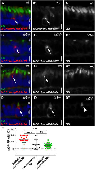
Outer segment development is rescued by constitutively active Rab8a. (A?A?) Cryosections of 4 dpf wt larvae with transient expression of WT mCherry-tagged Rab8a in cone PRs (tacp:mCherry-Rab8aWT) counterstained with DiO to mark OSs (green) and DAPI for nuclei. Note the punctate expression pattern of Rab8a concentrated in the inner segment space (arrow). (B?B?) A similar expression pattern is observed when expressing this transgene in ta3?/? PRs (arrow). (C?C?) Expression of a constitutively active form of mCherry-tagged Rab8a (tacp:mCherry-Rab8aCA) in wt cone PRs. (D?D?) ta3?/? PRs expressing constitutively active Rab8a develop OSs (arrowhead in D?). (E) Quantification of the proportion of 4 dpf ta3 mutant PRs expressing constitutively active Rab8a with OSs (red circles), ta3 mutant PRs expressing wild-type Rab8a with OSs (green squares) or ta3 mutant PRs without transgenic Rab8a expression with OSs (black triangles). Only the constitutively active form of Rab8a rescues OS formation in ta3 mutants (****p?<?0.0001, NS not significant, Student?s t-test, n?>?20 larvae expressing either transgene and 13 non-transgenic larvae). Each datapoint represents one individual larva. Bars are standard deviation. ONL outer nuclear layer, OS outer segment, S synapse. Scale bars: 3?Ám in all panels. |
|
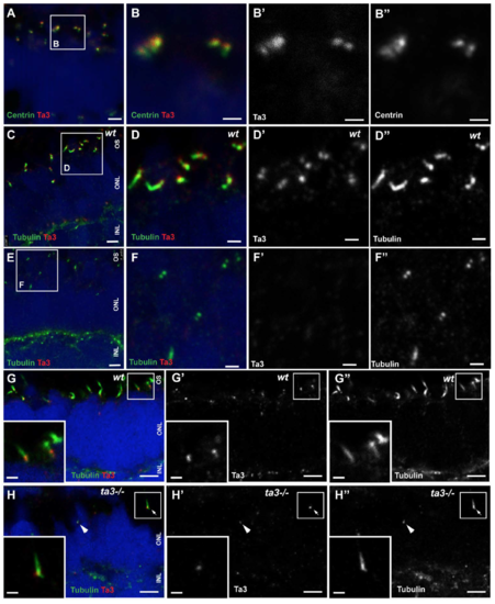
Ta3 protein localization time course in PRs (A-B'') 2 dpf retinal cryosections stained with anti-Centrin to mark BBs (green) and anti-Ta3 (red) show localization of Ta3 protein at the BB in wildtype (wt) larvae. (C-D'') 3 dpf cryosections of wt retinas stained with anti-acetylated tubulin and anti-Ta3 3 show localization of Ta3 at both mother and daughter centrioles. (E-F'') In contrast, Ta3 signal is abolished from the majority of BBs in 3dpf ta3 mutants. (G-G'') 4 dpf cryosections of wt retinas stained with anti-acetylated tubulin and anti-Ta3 show similar results as at 3 dpf. (H-H'') In ta3-/- PRs at 4 dpf, the Ta3 signal is mostly abolished (F'') except in a few isolated PRs (arrowhead and arrow in H-H'). Note that the two PRs still expressing Ta3 are also the only ones to have an extended axoneme (arrowhead and arrow in F''). The boxed area in A is shown in B-B'', the one in C is shown in D-D'' and the one in E is shown in F-F''. The insets in G-H'' represent the boxed areas in the corresponding images. Scale bars: 2.5 ?m in (A and C), 1 ?m in (B-B'', D-D'' and insets in E-F''), 4 ?m in (E-F''). |
|
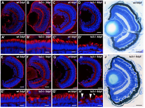
Time course of PR degeneration in ta3 mutants Retinal cryosections of wt and ta3-/- zebrafish at 3dpf (A-B'), 4dpf (C-D'), 5dpf (E-F') and 6 dpf (G-H'). Nuclei are counterstained with DAPI and membranes (including outer segments) are highlighted with BODIPY. Note the progressive cell shape loss and decreased numbers of outer segments in mutants. The arrow in H' indicates a PR with an OS, while the arrowheads point to PRs without OSs. (I-J) Histological sections through whole wt and ta3-/- eyes at 6dpf showing gaps in the PR cell layer, but also persistance of many PR cells. Scale bars: 30 ?m in (A-H) and (I-J), 4 ?m in A'-H'. |
|
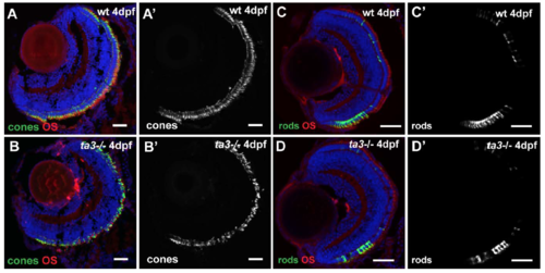
Differentiating ta3-/- PRs take on cone and rod specific cell fates (A-B) Retinal cryosections of 4 dpf wt and ta3-/- larvae stained with the zpr1 antibody indicating that mutant photoreceptors take on a cone-specific cell fate. (C-D') Likewise, ta3-/- photoreceptors take on a rod-specific fate as seen with the transgenic line tg(zfRH1-3.7B:EGFP) marking rod photoreceptors (PRs), seen here in retinal cryosections of 4dpf wt (C-C') and ta3 mutant (D-D') fish. Scale bars: 30 ?m in all panels. |
|
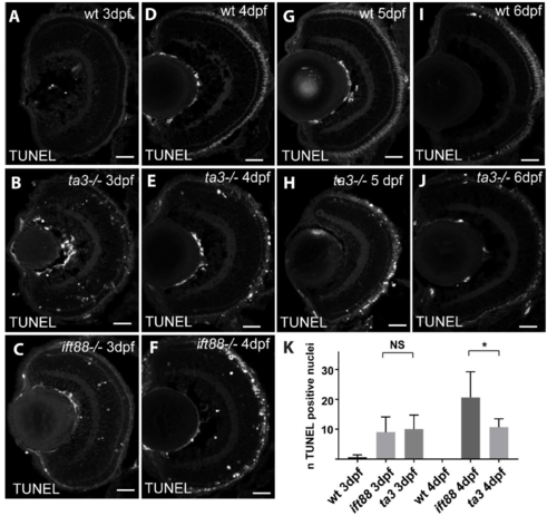
PR cell death in ta3 mutant retinae TUNEL staining on retinal cryosections at 3 dpf (A-C), 4 dpf (D-F), 5 dpf (G-H) and 6 dpf (I-J), in wildtype (A,D,G and I), talpid3 mutants (B,E,H and J) and ift88 mutants (C and F). Note the strong signal in ift88 mutants at 4 dpf and the comparatively lower amount of cell death in ta3 mutants at the same stage. (K) Quantification of cell death in wt, ift88 and ta3 mutants at 3 and 4dpf. The number of TUNEL-positive nuclei was counted on 5 ?m-thick confocal sections of entire retinal sections. Note the stable rate of cell death in ta3 mutants between 3 and 4dpf while ift88 mutants show an increase in cell death at 4dpf compared to 3dpf. Scale bars: 20 ?m in all panels. |
|
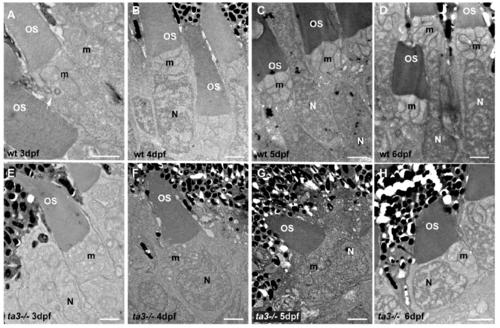
Progressive cell shape loss of ta3-/- PRs Transmission electron microscopy images of wt (A-D) and ta3-/- (E-H) PRs showing the progressive collapse of the normally apico-basally elongated cell shape of PRs. At 6 dpf, note the lack if inner segment space and the mitochondrial location next to the nucleus instead of its normal apical position (H). |
|
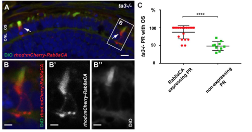
Rescue of OS development by expression of constitutively active Rab8a in rod PRs (A) Cryosection of 4 dpf zebrafish ta3-/- larva expressing constitutively active Rab8a in rod PRs (rhod:mCherry-Rab8aCA, red) and stained with DiO (green) to highlight membranes and OSs. Rod PRs expressing Rab8aCA develop OSs. (B) is a close-up image of the inset in (A). (B') shows only the red channel (Rab8aCA) and (B'') shows the green channel highlighting the presence of the OS. Scale bars are 4 ?m in A and 2 ?m in B-B''. (C) Quantification of OS development in rod PRs expressing Rab8aCA. Each red datapoint (circles) represents the percentage of PRs expressing the transgene that developed an OS in a single larva, while each green datapoint (squares) indicates the percentage of rod PRs ot expressing the transgene that developed OSs. |

