- Title
-
G9a and ZNF644 Physically Associate to Suppress Progenitor Gene Expression during Neurogenesis
- Authors
- Olsen, J.B., Wong, L., Deimling, S., Miles, A., Guo, H., Li, Y., Zhang, Z., Greenblatt, J.F., Emili, A., Tropepe, V.
- Source
- Full text @ Stem Cell Reports
|
Zebrafish znf644 Paralogs Are Specifically Expressed In and Regulate the Development of Retinal and Midbrain Progenitor Cells (A) Conservation of C2H2-like ZF motifs between human and zebrafish ZNF644 genes. (B) Representative lateral views, retinal cross-sections, and midbrain cross-sections of whole-mount in situ hybridization (WISH) assays monitoring znf644a or znf644b expression at 24 hpf (n > 15 in each group). Arrows denote the inner part of the central retinal epithelium. (C) Lateral views of g9a (20/32), znf644a (10/18), or znf644b (14/21) morphant embryos compared with WT (21/21). Dashed circles denote 2D area of the WT retina. Arrowheads denote the dorsal hindbrain region. (D) Quantitation (mean ± SD) of retinal 2D cross-sectional area (¼m2) of znf644a, znf644b, or g9a morphants relative to WT embryos. Error bars represent SD. *p < 0.005, Student′s t test. (E) RT-PCR assays monitoring unspliced (unspl) and spliced (spl) znf644a or znf644b transcripts at 24 hpf. |
|
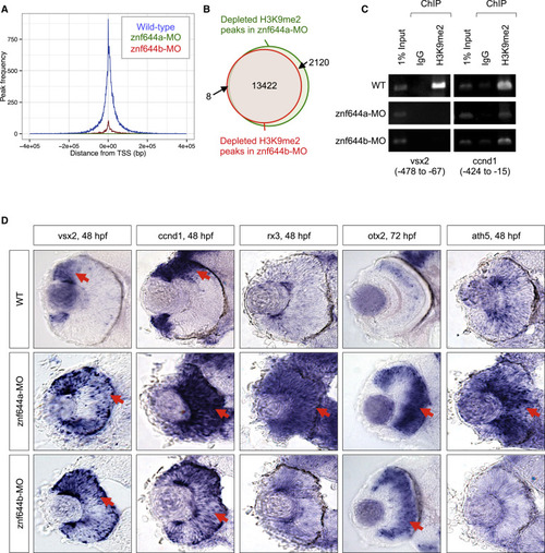
znf644a and znf644b Are Required for H3K9me2-Mediated Gene Silencing in the Forming Retina (A) Frequency and distance from TSS at which H3K9me2 peaks were identified in WT, znf644a, and znf644b morphants. (B) Venn diagram illustrating the number and overlap of H3K9me2 peaks at 48 hpf in znf644a or znf644b morphants or WT embryos. (C) ChIP-PCR assays monitoring H3K9me2 levels near the TSSs of vsx2 or ccnd1 genes at 48 hpf. (D) WISH assays monitoring expression of progenitor or proneural genes in WT, znf644a, or znf644b morphant retinas. Arrows highlight expression domains. EXPRESSION / LABELING:
PHENOTYPE:
|
|
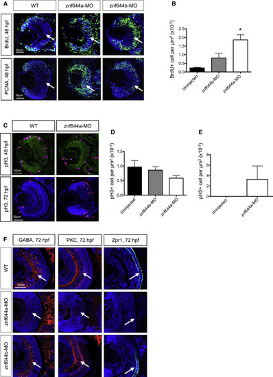
znf644a and znf644b Morphant Retinas Are Composed of Distinct Populations of Retinal Cells with Differing Characteristics (A) Immunostaining of retinal cross-sections monitoring BrdU incorporation and PCNA+ cells at 48 hpf in WT, znf644a, and znf644b morphants. (B) Quantitation of BrdU+ cells/¼m2 in central retina of WT, znf644a, or znf644b morphants at 48 hpf (n = 3 for each group). Error bars represent SD. p < 0.05, Student′s t test. (C?E) Immunostaining of pH3 expression in retinal cross-sections at 48 and 72 hpf in WT, and znf644a morphants (C). Quantitation of pH3+ cells/¼m2 in the central retina of WT, znf644a, or znf644b morphants at (D) 48 hpf (n = 5 for each group) and (E) 72 hpf (n = 2 for each group). Quantitation represented as mean ± SD. (F) Immunostaining monitoring marker protein expression in retinal cross-sections. At 72 hpf, amacrine cells express GABA, bipolar neurons express PKC, and cone photoreceptors express Zpr1. White arrows highlight populations of proliferative or differentiated cells. EXPRESSION / LABELING:
PHENOTYPE:
|
|
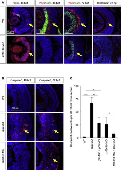
znf644a and znf644b Morphant Retinas Exhibit Distinct Cellular Defects (A) Immunostaining of retinal cross-sections of Vsx2 (red), Pax6 (red), Isl2b (green), or H3K9me2 (red) in WT or znf644a morphant embryos at indicated time points. Vsx2 expression at 48 hpf marks proliferating RPCs in the CMZ (gray arrows) and in a subpopulation of bipolar neurons in the central retina (yellow arrows). Pax6 expression marks amacrine cells and a subset of RGCs, and Isl2b expression marks a subset of RGCs. Differentiated retinal neurons with H3K9me2+ nuclei are highlighted. (B and C) Immunostaining monitoring cleaved Caspase3 (red) in retinal cross-sections from WT, g9a, or znf644b morphant embryos at 48 or 72 hpf (B). Yellow arrows highlight Caspase3+ apoptotic cells. (C) Quantitation of Caspase3+ cells in WT, znf644a morphant, or znf644b morphant retinas with or without co-injection of p53-MO at 72 hpf. Quantitation represented as mean ± SD (n=3 for each group). *p < 0.05, **p < 0.005, ***p < 0.0005, Student′s t test. EXPRESSION / LABELING:
PHENOTYPE:
|
|
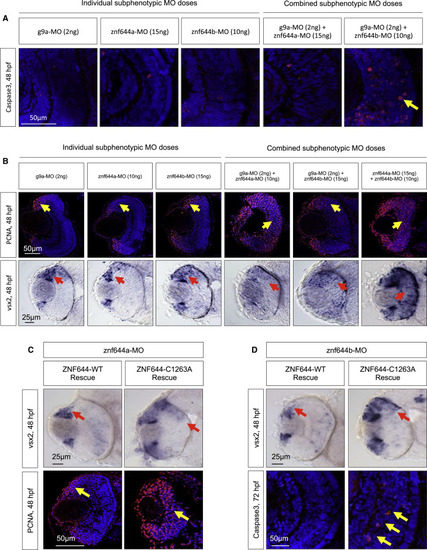
The Retinal Functions of znf644a and znf644b Are Dependent on Functional and Physical Interactions with g9a (A) Immunostaining of cleaved Caspase3 (red) in retinal cross-sections of embryos injected with individual or combined subphenotypic doses of g9a-MO, znf644a-MO, or znf644b-MO (n = 3 in each group). Yellow arrows highlight apoptotic cells. (B) (Top) Immunostaining of PCNA (n = 6 in each group) in retinal cross-sections from embryos injected with individual and combined subphenotypic doses of MOs (n = 6 in each group). Yellow arrows denote the position of the PCNA+ population. (Bottom) WISH assays monitoring the expression of vsx2 in retinal cross-sections of embryos injected with individual subphenotypic doses (n = 10 in each group) and combined subphenotypic co-injection of g9a-MO and znf644a-MO (n = 10), g9a-MO and znf644b-MO (n = 13), and znf644a-MO and znf644b-MO (n = 10). Red arrows denote the position of the vsx2 expressing cells. (C) (Top) WISH assays in retinal cross-sections monitoring vsx2 expression at 48 hpf in znf644a morphant embryos rescued by co-injection of either WT human ZNF644 mRNA (n = 12) or C1263A mutant mRNA (n = 9). (Bottom) Immunostaining of retinal cross-sections monitoring PCNA expression at 48 hpf showing rescue of znf644b morphants by co-injection of WT human ZNF644 mRNA (n = 3) but not C1263A mutants (n = 3). (D) (Top) WISH assays in retinal cross-sections at 48 hpf monitoring vsx2 expression from znf644b morphant embryos rescued by co-injection of human WT ZNF644 mRNA (n = 14) or C1263A ZNF644 mutant mRNA (n = 9). (Bottom) Immunostaining of retinal cross-sections monitoring cleaved Caspase3 at 72 hpf in znf644b morphant embryos rescued by co-injection of WT human ZNF644 mRNA (n = 3) or a C1263A mutant (n = 3). Red arrows highlight vsx2 expression domains, and yellow arrows highlight PCNA+ cell populations (C) or apoptotic cells (D). |
|
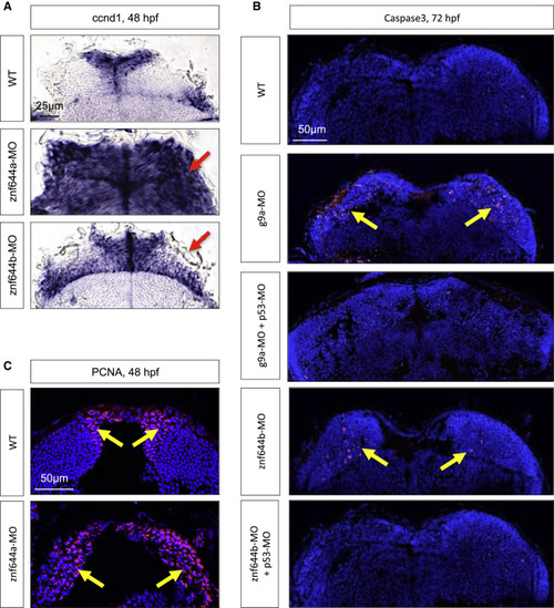
The Retinal Defects of g9a, znf644a, and znf644b Morphants Are Recapitulated in the Midbrain (A) WISH assays monitoring ccnd1 expression in WT, znf644a, or znf644b morphant midbrain cross-sections at 48 hpf. Arrows indicate mislocalized ccnd1 expression. (B) Immunostaining of cleaved Caspase3 in g9a or znf644b morphant midbrain cross-sections at 72 hpf and rescue by p53-MO (n = 3 in each group). Arrows denote the position of Caspase3+ cells. (C) Immunostaining of PCNA in WT or znf644a morphant midbrain cross-sections (n = 3 in each group). Arrows denote the position of PCNA+ cells. EXPRESSION / LABELING:
PHENOTYPE:
|
|
(A) Quantitative (q)PCR assays monitoring the relative expression level of ZNF644 mRNA from HEK293 cell lines stably expressing the indicated shRNA (n=3). Quantitation represented as mean ± SD. A non-silencing shRNA was used as a control. WISH assays monitoring the expression patterns of (B) znf644a and (C) znf644b. Both znf644a and znf644b are expressed maternally, and display widespread expression through the first 24hpf. At 48hpf expression is decreased, but still present in both anterior structures, and throughout the trunk. At each developmental stage no staining is apparent in sense strand control stained embryos. (D) RT-PCR assay monitoring the expression of znf644a, znf644b or β-actin cells from retinal extracts at the indicated time points. |
|
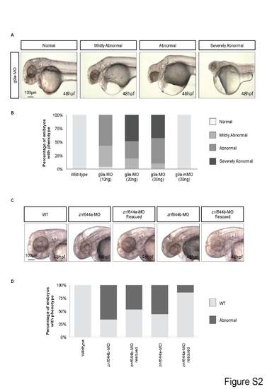
(A) Lateral views of WT or g9a morphant embryos illustrating the varying degrees of developmental defects observed. (B) Frequency at which the normal/abnormal morphologies are observed in WT or the indicated g9a-MO injected embryos. The mismatch control MO (g9a-mMO) is indicated. (C) Lateral views of WT, znf644a, or znf644b morphant embryos at 48 hpf as well as rescue assays in which the respective cognate mRNA were co-injected. (D) The frequency at which the WT or abnormal phenotypes were observed in indicated embryos. PHENOTYPE:
|
|
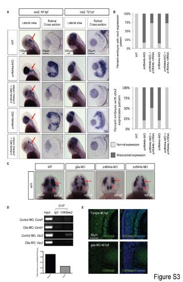
(A) WISH assays monitoring the expression of vsx2 at 48 hpf or otx2 at 72 hpf in lateral views or retinal cross-sections from WT, znf644a morphant or znf644b morphant embryos, as well as morphant embryos rescued by co-injection of the indicated mRNA. Red arrows indicate hindbrain neural cell populations that express vsx2. (B) Frequency at which the indicated embryos exhibited normal or mislocalized expression of vsx2 (top) or otx2 (bottom). (C) WISH assays monitoring the expression of lef1 at 48 hpf (dorsal views) in WT, g9a morphant, znf644a morphant, or znf644b morphant embryos. Red arrows point to lef1-positive midbrain regions, and red arrowheads point to lef1-expressing cells of the anterior hypothalamus. (D) ChIP-PCR assays monitoring the levels of H3K9me2 at the indicated positions near the TSS of vsx2 or ccnd1 genes (as in Figure 3C) at 48 hpf. An anti-H3K9me2 antibody and an isotype control (IgG) antibody were used for ChIP, followed by PCR amplification (35 cycles). Densitometry analysis revealed a ~50% reduction in the levels of H3K9me2 at the ccnd1 promoter, and complete loss of H3K9me2 levels at the vsx2 promoter in g9a morphant embryos. (E) Immunostaining with H3K9me2 antibody demonstrates a global reduction in nuclear expression in the g9a morphant retinas at 48 hpf compared to controls. EXPRESSION / LABELING:
PHENOTYPE:
|
|
Top: (A) WISH assays monitoring the expression of vsx2 or ccnd1 at 48 hpf in WT or g9a morphant retinal cross-sections. (B) Immunostaining assays monitoring BrdU- or pH3-postive cells at 48 hpf in WT or g9a morphant retinal cross-sections. (C) Immunostaining assays monitoring the expression of the indicated neuronal markers in g9a morphant retinal cross-sections at the indicated time points. Bottom: Blastula cell transplantation experiments (n=3 separate experiments) of donor morphant cells to wildtype host cells. All transplanted embryos were screened at 24 hpf and only those embryos with GFP+ contribution to the retina were further analyzed. (D-E) Only 2 out of 30 positively screened transplanted embryos contained GFP+ znf644a-MO cells within the retina at 72 hpf. (D) One embryo demonstrated a relatively large population of GFP+ morphant cells near the retinal periphery, which is more permissive for proliferation. Many donor cells at the periphery were also pH3+, and donor morphant cells located at the central retina were also proliferating ectopically (arrows). (E) The other embryo had very few cells in the center retina, but still showed evidence of ectopic proliferation (arrow). In both instances, host central retinal cells were not induced to proliferate. (F) Subphenotypic dose of GFP+ g9a-MO in donor cells does not lead to cell death after transplantation. (G) Subphenotypic dose of GFP+ znf644b-MO in donor cells does not lead to cell death after transplantation. (H) Combined subphenotypic doses of GFP+ g9a- MO and znf644a-MO results in donor cells undergoing cell death (caspase3+) that is confined to the donor cells. In each experiment, n>10 embryos were screened as GFP+ at 24 hpf, and n=3 were sectioned for analysis in each group at 48 hpf. Representative images of sections are shown. The WT ccnd1 (48 hpf) image in Figure S4A are re-used from Figure 3D; the WT BrdU (48 hpf) in Figure S4B is re-used from Figure 4A; the WT GABA, PKC and Zpr1 (72 hpf) images in Figure S4C are re-used from Figure 4F; and the WT vsx2 (48 hpf) images in Figure S4C are re-used from Figure 5A. EXPRESSION / LABELING:
PHENOTYPE:
|
|
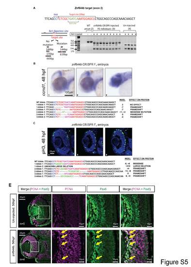
Top: CRISPR-Cas9 targeting of the znf644b gene show multiple mutated alleles in the F1 generation that correlates with cellular defects in the retina. (A) PCR and restriction digest diagnostic on genomic DNA extracted from individual F0 embryos injected with Cas9 mRNA and sgRNA targeting against exon 2 of znf644b. The PCR fragment contains two Bc1I digestion sites, one of which is located within the sgRNAtargeting site. Digestion of wild-type sequences results in three fragments, 449bp, 170bp and 144bp, whereas CRISPR-mediated insertion/deletion mutations result in loss of the digestion site in the target site, resulting in an additional 619bp fragment. (B, top) WISH assays monitoring the expression of ccnd1 in F1 CRISPR embryos at 48hpf. One F1 embryo heterozygous for a frameshift mutation and was phenotypically normal for brain and retinal growth as well as ccnd1 expression at 48 hpf (embryo#1). A separate embryo with two mutant (frameshift) alleles showed significantly reduced brain and retinal growth and elevated expression of ccnd1 (embryo#3), which was reminiscent of the znf644b morphant phenotype. (C, top) Immunostaining assay monitoring pH3-positive cells in retinal cross-sections from F1 CRISPR embryos at 48hpf. One embryo had two znf644b mutant alleles, one of which was predicted to be a very large deletion likely resulting in a non-functional protein, while the other allele had a missense mutation leading to a predicted substitution of only two amino acids. The overall phenotype of this embryo was comparable to WT, suggesting that this embryo was functionally heterozygous (embryo#1). Other embryos genotyped as heteroallelic mutants displayed phenotypes that were similar to the morphants, such as a reduced retinal size and persistent pH3+ cells in the central retina at 48 hpf (embryo#2: 11.67 ± 4.16; embryo #3: 12.67 ± 2.08 compared to WT: 3.8 ± 0.5, n=9). (B & C, bottom) The corresponding sequencing data for both alleles of the embryos analyzed, with the predicted effect on the protein function. Black letters represent unchanged bases; Blue letters represent the PAM sequence; Red letters represent the target sequence; Pink letters represent the Bc1I digestion site; Dashes represent deletions; Green letters represent inserted bases. Bottom: Expression overlap of cell cycle regulator PCNA and the retinal differentiation marker Pax6 in the znf644b morphant retina. At 56 hpf, which coincides with the onset of widespread cell death of central retinal cells in znf644b morphants, PCNA can be found to be expressed in differentiated amacrine and ganglion cells (Pax6-postive) in the znf644b morphant retina, even though this population of neurons is reduced overall. |
|
Interaction of g9a and znf644 genes in regulating progenitor cell cycle. (A) WISH assays monitoring the expression of ccnd1 in (top) midbrain or (bottom) retinal cross-sections at 48 hpf from WT embryos or embryos injected with the indicated combined subthreshold MO doses. (B) Immunostaining assays monitoring pH3 levels or BrdU incorporation at 48 hpf from WT or embryos injected with the indicated combined subthreshold MO doses. (C) Rescue experiments in which mRNA encoding WT human (ZNF644) or mutant version lacking G9a/GLP binding capability (C1263A) is coinjected with znf644a-MO (left) or znf644b-MO (right). Vsx2 expression was assessed by WISH at 48 hpf, and PCNA, pH3, and Zpr1 levels (n=3 in each group) were assessed by immunostaining and confocal microscopy at the indicated time points. |


