
Fig. 5
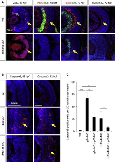
znf644a and znf644b Morphant Retinas Exhibit Distinct Cellular Defects (A) Immunostaining of retinal cross-sections of Vsx2 (red), Pax6 (red), Isl2b (green), or H3K9me2 (red) in WT or znf644a morphant embryos at indicated time points. Vsx2 expression at 48 hpf marks proliferating RPCs in the CMZ (gray arrows) and in a subpopulation of bipolar neurons in the central retina (yellow arrows). Pax6 expression marks amacrine cells and a subset of RGCs, and Isl2b expression marks a subset of RGCs. Differentiated retinal neurons with H3K9me2+ nuclei are highlighted. (B and C) Immunostaining monitoring cleaved Caspase3 (red) in retinal cross-sections from WT, g9a, or znf644b morphant embryos at 48 or 72 hpf (B). Yellow arrows highlight Caspase3+ apoptotic cells. (C) Quantitation of Caspase3+ cells in WT, znf644a morphant, or znf644b morphant retinas with or without co-injection of p53-MO at 72 hpf. Quantitation represented as mean � SD (n=3 for each group). *p < 0.05, **p < 0.005, ***p < 0.0005, Student′s t test. |
| Gene: | |
|---|---|
| Antibodies: | |
| Fish: | |
| Knockdown Reagents: | |
| Anatomical Terms: | |
| Stage Range: | Long-pec to Protruding-mouth |
| Fish: | |
|---|---|
| Knockdown Reagents: | |
| Observed In: | |
| Stage Range: | Long-pec to Protruding-mouth |