- Title
-
Arf6 and the 5'phosphatase of synaptojanin 1 regulate autophagy in cone photoreceptors
- Authors
- George, A.A., Hayden, S., Stanton, G.R., Brockerhoff, S.E.
- Source
- Full text @ Bioessays
|
Abnormalities in nrca14 cones are specific to late endosomes and autophagosomes and appear early in development. Images of fixed wild type (WT) and nrca14 retinas from A: Tg(TαCP : GFP-map1lc3b) and B: Tg(TαCP : GFP-rab7) larvae at 3 and 4 days post-fertilization (dpf). C: nrca14 cones contain more LC3 positive puncta than WT cones by 3 dpf (compare left panels in A). B: Abnormally enlarged Rab7 structures are present in nrca14 cones by 3 dpf. D: Images of fixed WT and nrca14 retinas from 5 dpf Tg(TαCP : GFP-Rab5a) larvae. There is no difference in the appearance or number of Rab5a positive early endosomes between WT and nrca14. Scale bar = 2 µm in all images. Graph C shows average LC3 puncta per cell at 3 or 4 dpf, error bars are standard error of mean (SEM). n = 8 WT larvae and seven nrca14 larvae on 3 dpf and n = 8 WT larvae and nine nrca14 larvae on 4 dpf. (*p-value < 0.05, ***p-value < 0.001 as assessed by Mann-Whitney test). Graph E shows average Rab5a puncta per cell at 5 dpf, error bars are SEM. n = 4 WT larvae and four nrca14 larvae. (ns = p-value > 0.05 as assessed by Mann?Whitney test). EXPRESSION / LABELING:
PHENOTYPE:
|
|
Maturation of autophagosomes is blocked in nrca14 cones. Expression of the tandem mCherry-GFP-LC3 probe in cones of wild type (WT) and nrca14 5 days post-fertilization larvae. A: In WT photoreceptors, autolysosomes (magenta-only puncta) were visible (top panel). A: Fewer magenta-only puncta were visible in nrca14 cones (bottom panel), indicating that the LC3 positive autophagosomes had not fused with an acidic compartment. B: Shows enlargement of areas shown in boxes in A. White arrow heads indicate GFP+, mCherry+ autophagosomes, magenta arrowheads point to GFP-, mCherry+ autolysosomes. Scale bar = 2 µm in A. Graph in C shows average number of autophagosomes (GFP+, mCherry+) and autolysosomes (GFP-, mCherry+) per 20 µm3. Error bars are SEM and n = 7 WT larvae and six nrca14 larvae. (***p-value < 0.001, *p-value < 0.05 respectively as assessed by Mann-Whitney test). |
|
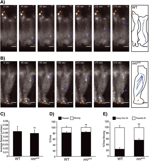
Mobility of autophagosomes is not reduced in nrca14 cones. Live time-lapse images of Tg(TαCP : GFP-map1lc3b) 5 days post-fertilization zebrafish larvae show that autophagosomes are highly mobile in both wild type (WT) and nrca14 cones. Images were acquired in 5 second frame intervals. Selected time points are shown for A: WT and B: nrca14 cones. A schematic showing the outline of the cell(s) and movement of two representative LC3 positive particles are shown for each genotype. Puncta in nrca14 cones exhibit similar movements to those found in WT. C: The average speed and D: relative amount of time LC3 positive vesicles were mobile is the same in WT and nrca14 cells. E: In nrca14 cells, LC3 vesicles spent significantly more time moving away from the inner segment (IS) than in WT cells. Error bars are SEM and n = 15 vesicles from three WT larvae and 15 vesicles from three nrca14 larvae. (ns = p-value > 0.05, **p-value < 0.01 as assessed by Mann?Whitney test). Scale bar = 2 µm. EXPRESSION / LABELING:
|
|
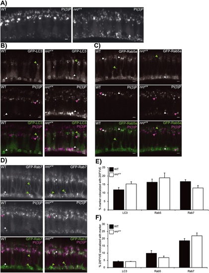
nrca14 cone photoreceptors have an increased number of PI(3)P positive puncta, but no change in distribution of PI(3)P. A:. PI(3)P was visualized in wild type (WT) and nrca14 Tg(TαCP : YFP-2XFYVE) transgenic larvae at 5 days post-fertilization. The PI(3)P probe localizes to punctate structures in the inner segment and synaptic terminals of both WT and nrca14 cones. More PI(3)P positive puncta are visible at the synaptic terminals of nrca14 cones relative to WT cones. PI(3)P was marked using Tg(TαCP : tagRFPt-2XFYVE) in 5 days post-fertilization cones expressing B: GFP-LC3, C: GFP-Rab5a or D: GFP-Rab7. Examples of puncta with only GFP signal, only tagRFPt signal or both signals are denoted by green, magenta or white arrowheads, respectively. E: Percent colocalization of indicated marker with 2XFYVE. F: Percent colocalization of the 2XFYVE probe with indicated marker. E and F: We found no significant difference in the percent colocalization of either LC3, Rab5a or Rab7 with 2XFYVE between WT and nrca14 cones. Error bars are SEM and n = 7-8 WT larvae and 4-6 nrca14 larvae. (ns = p-value > 0.05 as assessed by Mann?Whitney test). Scale bar = 2 µm. EXPRESSION / LABELING:
|
|
The distribution of PI(3,5)P2 positive late endosomal compartments is the same in wild type (WT) and nrca14 cells. A; The PI(3,5)P2 probe ML1NX2 localizes to small, punctate structures in the inner segments of WT and nrca14 cone photoreceptors in 5 days post-fertilization Tg(TαCP : mCherry-ML1NX2) transgenic larvae. B: Some PI(3,5)P2 puncta overlap with LC3 positive puncta in nrca14 but not WT cones, agreeing with the decrease in acidic LC3-positive structures observed in Fig. 2. C: The PI(3,5)P2 partially overlaps with Rab7 puncta in both WT and nrca14 cone photoreceptors, but is not found on abnormal Rab7 structures in nrca14 cones. Scale bar = 2 µm in all images. EXPRESSION / LABELING:
|
|
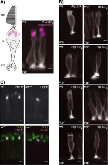
PI(4,5)P2 and PI(4)P distributions are not altered in nrca14 cones. A and B: The PI(4,5)P2 probe GFP-PLCδ-PH was expressed in photoreceptors using the crx promoter. A: In wild type (WT) cells at 5 days post-fertilization (dpf), this probe localized to the plasma membrane and concentrated at synapses, but did not extend above the mitochondria (magenta) into the outer segment (OS). C: The PLC´-PH probe showed the same distribution in nrca14 photoreceptors from 3 to 6 dpf. PI(4)P was visualized with the probe RFP-FAPP1-PH. In both WT and nrca14 photoreceptors at 5 dpf, the RFP-FAPP1-PH probe (magenta) overlapped with the medial Golgi marker Man2a-GFP (green). There was no difference in the subcellular distribution of PI(4)P between WT and nrca14 photoreceptors. Scale bar = 2 µm in all images. IS, inner segment. |
|
5′phosphatase activity of SynJ1 is required to rescue late endosome and autophagy phenotypes in nrca14 cones. Fixed sections of 5 days post-fertilization nrca14 A: Tg(TαCP : GFP-map1lc3b) and B: Tg(TαCP : GFP-rab7) larvae transiently expressing synaptojanin 1 (SynJ1)constructs (SJCs) in cones. Wild type SynJ1 was able to significantly rescue both the late endosome and autophagosome abnormalities in nrca14 cones. SynJ1 lacking SacI catalytic activity (C385S) also significantly rescued the late endosome and autophagosome defects. In contrast, the D732A construct was not able to rescue either the late endosome or autophagosome phenotypes. Arrows point to SJC-expressing cells (magenta in merge). Scale bar = 2 µm in all images. Results were quantified by counting the number of total C: LC3 puncta or D: Rab7 synaptic puncta in the SJC-expressing cells and normalized to the number of puncta in the non-SJC expressing cells in the same nrca14 larvae. Graph in C shows average normalized LC3 puncta per cell, and graph in D shows average normalized synaptic Rab7 puncta per cell. Error bars are SEM and n ≥ 50 cells for all conditions (exact n shown on graph). The rescue of both the total LC3 puncta and Rab7 synaptic puncta was significant for the wild type and C385S SJCs, but not for the D732A SJC (***p-value < 0.001 by one-way analysis of variance followed by Dunnett′s multiple comparison correction). |
|
Altered Arf6a activity can rescue autophagy phenotype in nrca14 cones. Fixed sections of 5 days post-fertilization A: wild type (WT) and B: nrca14 Tg(TαCP : GFP-map1lc3b) larvae transiently expressing Arf6a-tagRFPt construct in cones. WT Arf6a did not significantly affect the number of autophagosome in WT or nrca14 cones. Constitutively active Arf6a Q67L significantly rescued autophagosome defects nrca14 cones. Dominant negative guanosine diphosphate (GDP)-bound Arf6a T44N rescued the autophagy phenotype in nrca14 cones and decreased the number of autophagosomes in WT cones. Dominant negative phospholipase D-binding deficient Arf6a N48R also significantly rescued the autophagy defect in nrca14 cones. Arrowheads point to Arf6a expressing cells (magenta in merge). Scale bar = 2 µm in all images. Results were quantified by counting the number of total LC3 puncta in the Arf6a expressing cells and background corrected using the number of puncta in the non-Arf6a expressing cells in the same larvae. Graph in C shows average corrected LC3 puncta per cell. Error bars are SEM and n ≥ 50 cells for all conditions (exact n shown on graph) (*p-value < 0.05, ***p-value < 0.001 relative to cells not expressing Arf6a in nrca14, #p-value < 0.05 in WT, by one-way analysis of variance followed by Dunnett′s multiple comparison correction). EXPRESSION / LABELING:
|
|
Constitutively active Arf6a Q67L rescues late endosome phenotype in nrca14 cones. Fixed sections of 5 days post-fertilization A: wild type (WT) and B: nrca14 Tg(TαCP : GFP-rab7) larvae transiently expressing Arf6a-tagRFPt construct in cones. WT and dominant negative Arf6a did not significantly affect the number of synaptic Rab7 puncta in WT or nrca14 cones. Constitutively active Arf6a Q67L significantly rescued the synaptic Rab7 puncta in nrca14 cones. Arf6a Q67L significantly increased the number of synaptic Rab7 puncta in WT cones. Arrowheads point to Arf6a-expressing cells (magenta in merge), arrows point to Arf6a-expresssing synaptic terminals. Scale bar = 2 µm in all images. Results were quantified by counting the number of synaptic Rab7 puncta in the Arf6a expressing cells and background corrected using the number of puncta in the non-Arf6a expressing cells in the same larvae. Graph in C shows average corrected synaptic Rab7 puncta per cell. Error bars are SEM and n ≥ 50 cells for all conditions (exact n shown on graph) (* p-value < 0.05,*** p-value < 0.001 in nrca14, #p-value < 0.05 in WT relative to cells not expressing Arf6a by one-way analysis of variance followed by Dunnett′s multiple comparison correction). EXPRESSION / LABELING:
|
|
|
|
|
|
|


