- Title
-
Zebrafish Wnt9b Patterns the First Pharyngeal Arch into D-I-V Domains and Promotes Anterior-Medial Outgrowth
- Authors
- Jackson, H.W., Prakash, D., Litaker, M., Ferreira, T., Jezewski, P.A.
- Source
- Full text @ Am. J. of Molec. Bio.
|
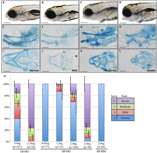
Gross morphological phenotypes of 5dpf zebrafish after Wnt9b knockdown. 5dpf live (A)-(D) and Alcian blue stained (E)-(L) embryos, anterior to left, lateral view (A)-(H) and ventral view (I)-(L). Control embryos, injected with the five base mismatched splice blocking morpholino (SB-MM) are shown within the left most column (A), (E), (I) and are indistinguishable from uninjected controls. Splice blocking morpholino (SB-MO) injected embryos were scored as Mild (B), (F), (J), Moderate (C), (G), (K) or Severe (D), (H), (L) using an objective scoring method (see Supplemental Figure 1. for details) and are shown in subsequent columns. (M) Quantification of the proportion of embryos within each category. Translation blocking (TB-MO), splice blocking (SB-MO) and mismatch control (SB-MM) injected embryos were categorically scored at 5dpf. Injection dose is indicated in ng, and the total number (n) of embryos scored is listed. Note that 5.3 ng TB-MO and 2.6 ng SB-MO produce comparable proportions of affected embryos. Error bars are standard error of means of three biological replicates. Size bars in (A)-(L) are 250 um. PHENOTYPE:
|
|
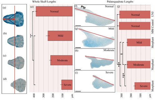
Dissected morphology of Wnt9b morphants reveals significant skull and palatoquadrate (PQ) foreshortening along the anterior-posterior (A-P) axis. Alcian blue whole-skulls (a)-(d) or dissected PQs (f)-(i) were flat mounted and digitally measured. Normal (A), (F) MM control and mild (b), (g), moderate (c)-(h) and severe (d), (i) skeletal elements from SB-MO injected embryos. (a)-(d) Red line aligns these skulls along the common interhyal joint, with digital measurements performed perpendicular to this line to the anterior-most tip of the Meckel?s cartilage (red arrows). Note that the anterior tip of the notochords are also aligned. (e) Bar chart showing mean skull lengths per category. All morphant skulls grouped together were significantly shorter than controls. (f) (i) Red line represents the digital measurement of the PQ along the A-P axis. Size bars in (f)-(i) are 40 Ám. (j) Bar chart showing mean PQ lengths per category, and morphant PQs are significantly shorter than controls. Note that uninjected control and MM control PQs are not significantly different. +p = 0.071, ***p < 0.001. 1-way ANOVA analysis, Tukey?s HSD test of means. Ptp = pterygoid process. PHENOTYPE:
|

ZFIN is incorporating published figure images and captions as part of an ongoing project. Figures from some publications have not yet been curated, or are not available for display because of copyright restrictions. |
|
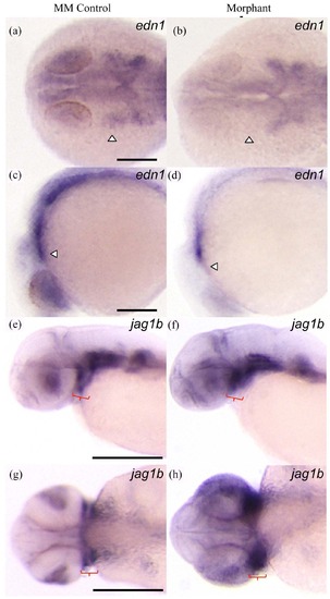
Wnt9b morphants exhibit moderate reduction of edn1 expression and the expected expansion in jag1b expression in the pharyngeal arches. (a)-(d) Anterior to right, top row = dorsal view, bottom row = lateral view. MM control (n = 8) (a), (c) and morphant (n = 8) (b, d) embryos were aged to 24hpf and stained with edn1 antisense riboprobe. 63% of morphant embryos exhibited reduced edn1 expression in the pharyngeal arches compared to controls. (e)-(f) Top row = lateral view, bottom row = anterior view. jag1b expression in 48hpf aged MM control (n = 14) (e), (g) and morphant (n = 10) (f)-(h) embryos. 100% of morphant embryos exhibited expanded jag1b expression in the pharyngeal arches compared to controls. White arrowheads indicate PA1. Red brackets indicate PA1. Size bars = 100 Ám. |
|
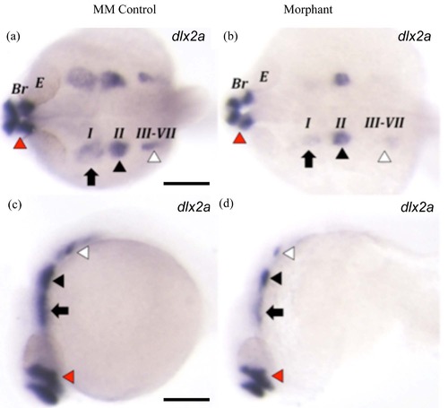
Wnt9b morphants exhibit severely reduced dlx2a expression in pharyngeal arches 1 and 3-7. (a)-(d) Anterior to right, top row = dorsal view, bottom row = lateral view. MM control (n = 17) (a), (c) and morphant (n = 20) (b), (d) embryos were aged to 24hpf and stained with dlx2a antisense riboprobe. 60% of morphant embryos exhibited reduced dlx2a expression in the pharyngeal arches compared to controls. Red arrowheads indicate forebrain. Black arrows indicate PA1, black arrowheads indicate PA2, white arrowheads indicate PA3-7. Note that dlx2a expression is mostly reduced in PA1 and PA3-7. Br = brain, E = eye. Size bars = 100 Ám. |
|
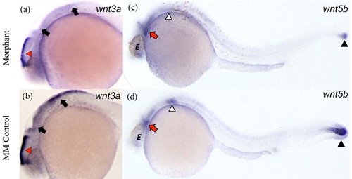
Wnt9b morphants exhibit disrupted Wnt3a and Wnt5b expression domains. Anterior to left. Left column is Wnt3a expression, right column is Wnt5b expression. (a) MM control (n = 14) and (b) morphant embryos (n = 14) were aged to 28hpf and stained with Wnt3a riboprobe. 71% of morphant embryos exhibited expanded Wnt3a expression in the brain compared to controls. (c) MM control (n = 10) and (d) morphant embryos (n = 9) were aged to 28hpf and stained with Wnt5b riboprobe. 100% of morphant embryos exhibited expanded Wnt5b expression in the tail with reduced Wnt5b expression in the pharyngeal arches compared to controls. (a), (b) Red arrowheads indicate brain expression, black arrows indicate rhombic lip expression. (c), (d) Red arrows indicate the PAs, white arrowheads indicate the fin buds, black arrowheads indicate the tail. |
|
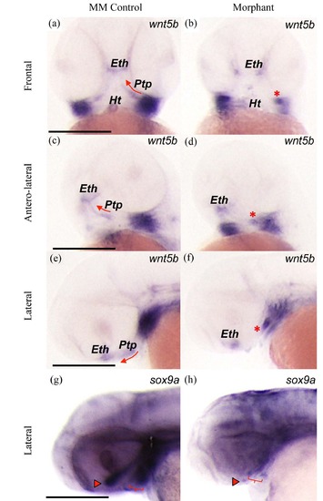
Wnt9b morphants display disrupted Wnt5b and sox9a expression domains in the developing upper jaw. MM control (n = 11) (left column) and morphant (n = 13) (right column) embryos were aged to 48hpf and stained with Wnt5b riboprobe (a)-(f) or sox9a riboprobe (g), (h). 85% of morphant embryos exhibited disrupted Wnt5b expression in the pharyngeal arches compared to controls. 73% of morphant embryos exhibited reduced sox9a expression in the pharyngeal arches compared to controls. Frontal views (a), (b), antero-lateral views (c), (d), and lateral views (e)-(h) with anterior to the right. Red arrows trace the linear outgrowth of the pterygoid process. Red asterisks indicate clumped pterygoid process cells. Red arrowheads indicate the ethmoid. Red brackets indicate the first pharyngeal arch precartilage condensations. E = eye, Eth = ethmoid, Ht = heart tube, Ptp = pterygoid process. Size bars = 100 Ám. |
|
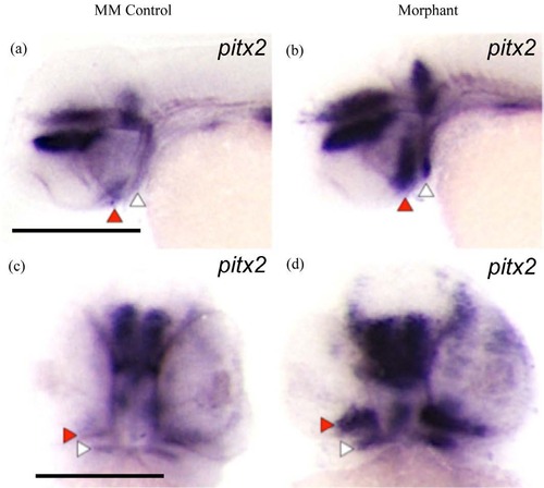
Wnt9b morphants exhibit expanded pitx2 expression domains in the developing mouth. Top row = anterior to left, bottom row = anterior to front. MM control (n = 8) (a), (c) and morphant (n = 11) (b), (d) embryos were aged to 48hpf and stained with pitx2 riboprobe. 82% of morphant embryos exhibit expanded pitx2 expression within medial oral regions compared to controls. Red arrowheads indicate upper jaw, white arrowheads indicate lower jaw. Size bars = 100 Ám. |
|
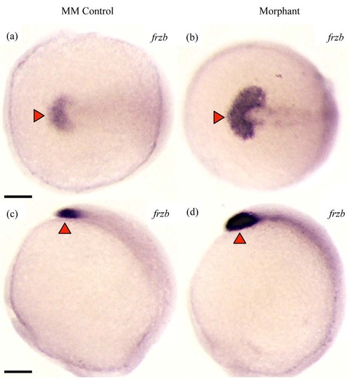
Wnt9b morphants exhibit expanded frzb expression in the polster. Top row = dorsal view, bottom row = lateral view, anterior to left. MM control (n = 6) (a), (c) and morphant (n = 15) (b), (d) embryos were aged to 13hpf and stained with frzb riboprobe. 93% of morphant embryos exhibit expanded frzb expression compared to controls. Red arrowheads indicate the polster. Note that morphants have upregulated and expanded expression of frzb. Size bars = 100 Ám. EXPRESSION / LABELING:
PHENOTYPE:
|
|
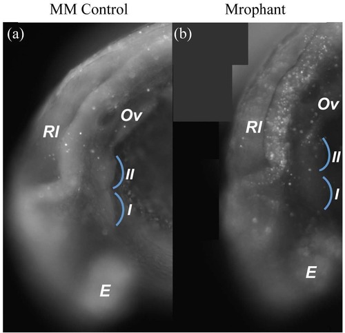
Wnt9b morphants do not display increased apoptosis in the first pharyngeal arch. (a) (b) Lateral Views. (a) MM control and (b) morphant embryos were aged to 26hpf and subjected to acridine orange staining to assay cell death. Blue arcs indicate PA1&2. Note that apoptotic cells within the PAs are not increased in morphants. Apoptotic cells within dorsal neural structures are increased in morphants. Br = brain, E = eye, Ov = otic vesicle, Rl = rhombic lips. |
|
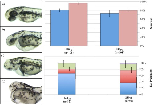
Wnt9b overexpression reduces dorsal/neural tissues. (a)-(d) 1-cell aged embryos were injected with either 140 pg or 280 pg of synthetic Wnt9b mRNA and monitored for survival (e) and 48hpf eye phenotypes (f). Live embryos scored for (a) normal eyes, (b) small eyes, (c) cyclopia, and (d) no eye phenotypes that are represented in (f). (b) Blue columns are the 0 - 1dpf survival, red columns are the 1 - 2dpf survival. (f) Distribution of eye phenotypes of Wnt9b overexpressing embryos. Purple = no eyes, green = cyclopic, red = small eyes, blue = normal eyes. |

