- Title
-
Genetic Interaction between pku300 and fbn2b Controls Endocardial Cell Proliferation and Valve Development in Zebrafish
- Authors
- Wang, X., Yu, Q., Wu, Q., Bu, Y., Changm, N.N., Yan, S., Zhou, X.H., Zhu, X., and Xiong, J.W.
- Source
- Full text @ J. Cell Sci.
|
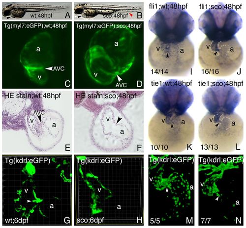
Endocardial and cardiac valve defects were found in scote382 mutant embryos. (A,B) Brightfield views of live wild-type (A) and scote382 mutant (B) embryos at 48hpf. Note pericardial edema and heart abnormality (black arrowhead), as well as abnormal vessels in the caudal trunk (red arrowhead). (C,D) Confocal images of Tg(myl7:eGFP) transgenic wild-type (C) and scote382 mutant (D) hearts at 48hpf. Note lack of constricted AVC and dilated heart in scote382 mutant (arrowhead). (E,F) Hematoxylin-eosin staining of wild-type (E) and scote382 mutant (F) hearts at 48hpf on transverse JB4 sections. Note lack of AVC constriction (arrowhead) in scote382 mutant heart. (G,H) Valve leaflets (arrowhead) were labeled and detected by the Tg(kdrl:eGFP) transgene in wild-type (G), but not in scote382 mutant (H) embryos at 6dpf on vibrotome sections across the heart. (I?N) Endocardial markers fli1 (I,J) and tie1 (K,L) were expressed in wild-type (I,K) and scote382 mutant (J,L) embryos; and Tg(kdrl:eGFP) labeled the endocardium in wild type (M) and scote382 mutant (N) embryos at 48hpf. Note that endocardial genes (fli1, tie1 and flk1) were reduced in the atrium of scote382 mutants (J,L,N). Arrows point to the AVC. a, atrium; v, ventricle. |
|
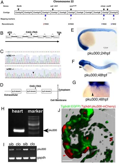
Positional cloning of pku300 from the scote382 locus in zebrafish. (A) The scote382 locus was mapped on linkage group 22 by bulk segregation analysis with microsatellite markers. Note that the scote382 locus, which contains calr, trh1, pku300 and cherp genes, was determined between markers 6 and 9 by genotyping 1632 informative meiosis using SLP and SNP methods. A G3935T missense mutation of fbn2b is reported to be responsible for the scote382 locus (Mellman et al., 2012), but fbn2b is located outside of the genetic interval in our mapping cross DNA panels. fbn2b is about 570kb away from pku300. (B) pku300 consists of 6 exons and encodes a 540-amino-acid protein. Note that a C946T mutation in exon 3 of pku300 led to a premature stop code (TAG) in the scote382 allele (arrowhead). (C) Sequencing traces of wild-type and homozygous scote382 mutant embryos. Note a single base pair change from C to T in the scote382 allele (arrowhead). (D) Pku300 protein was predicted to contain three extracellular IgG domains, a transmembrane domain and a short intracellular tail. (E?G) pku300 was broadly expressed, including in the heart (arrowhead) during early embryogenesis. Staged wild-type 24 (E), 48 (F) and 48hpf (G) hearts were subjected to in situ hybridization with anti-sense pku300 riboprobe. (H) pku300 was expressed in day-2 Tg(myl7:eGFP) embryonic hearts by RT-PCR. (I) pku300 was reduced in cloche (clo) mutant embryos that have no endocardial cells. RNA was prepared from siblings (sib) and clo mutant embryos (clo) from heterozygous clochem39 crosses. (J) Pku300-mCherry fusion proteins were transiently expressed in the heart of a Tg(kdrl:EGFP) transgenic embryo. Note the red Pku300-mCherry fusion protein localization on cell membranes (arrowhead), which was not overlapped with cytoplasmic localization of EGFP. Pku300-mCherry was driven by the zebrafish kdrl promoter. a, atrium; v, ventricle. Scale bar: 30μm. |
|
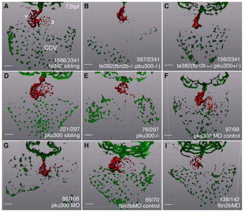
Mutations of both pku300 and fbn2b contributed to scote382 mutant phenotypes. The endocardium in the atrium, ventricle and CCV were labeled by the Tg(kdrl:EGFP) transgene. The endocardium was projected by red pseudocolor with Imaris software. (A-C) 2341 offspring from heterozygous scote382 mutant crosses produced 25.5% mutants that had defects in the atrial endocardium and CCV (B), 6.7% mutants that had only reduced atrial endocardium (C), and 67.8% siblings that were phenotypically normal (A), suggesting more than one gene mutated in the scote382 locus. Sequencing confirmed that the mutants (group II) with defects in the heart (endocardium and CCV) and tail (caudal vein) (supplementary material Fig. S3) were fbn2b-/- and pku300-/- (B), and the mutants (group I) with defects only in the heart (endocardium) were fbn2b+/- and pku300+/- (C). (D?F) 297 offspring from heterozygous scopku300 mutant crosses produced 25.5% mutants with defects in the atrial endocardium and CCV (E) and 74.6% normal siblings (D), which was supported by morpholino knockdown of pku300 with 9.6ng pku300MO (F). (G?I) A fbn2bMO (1.6ng) morphant showed reduced atrial endocardium but had normal CCV (I), compared with the control (H) and a pku300 morphant (G). a, atrium; v, ventricle. Scale bars: 50μm. EXPRESSION / LABELING:
PHENOTYPE:
|
|
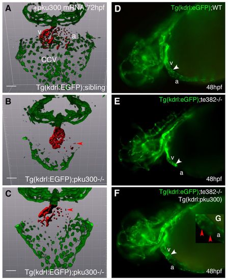
Both pku300 mRNA and transgenic expression of pku300 in the endocardium rescued the respective mutant phenotypes of scopku300 or scote382. (A-C) The endocardium in the atrium, ventricle and CCV were labeled by Tg(kdrl:eGFP). The endocardium was projected as red pseudocolor with Imaris software. Compared with a wild-type sibling (A), pku300 mRNA rescued the atrial endocardium and CCV of a scopku300 mutant embryo (C). Nine homozygous scopku300 mutants out of 182 offspring from heterozygous scopku300 crosses showed little or no rescue in the atrial endocardium and CCV (B). Wild-type sibling (A) and homozygous scopku300 mutants (B,C) were confirmed by sequencing. Red arrowheads point to the endocardium. (D-G) The endocardium was labeled by Tg(kdrl:eGFP) in the atrium and ventricle of wild-type embryos (D). Note little endocardium in the atrium of scote382 mutant Tg(kdrl:eGFP) embryos (E), which were partially rescued by overexpressing pku300 using Tg(kdrl:pku300) transgene (F). (G) Higher magnification of the atrium of panel F shows the rescued atrial endocardial cells (red arrowheads). The atrium and ventricle are demarcated with white arrowheads (D-F). a, atrium; v, ventricle; WT, wild type. Scale bars: 50μm. |
|
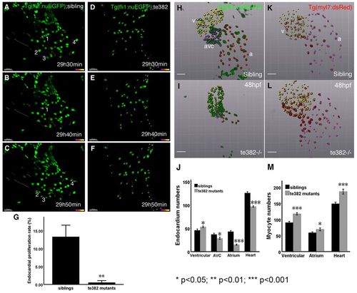
Atrial Tg(fli1:nuEGFP) endocardial numbers were reduced while ventricular cardiomyocyte numbers were increased in scote382 mutant embryos. (A?F) Endocardial nuclei were labeled by Tg(fli1:nuEGFP) in wild-type (wt) (A?C) and scote382 mutant (D?F) embryos at 29hours 30minutes, 29hours 40minutes and 29hours 50minutes post fertilization. A representative set of time-lapse images showed one divided cell from 29hours 30minutes to 29hours 40minutes (1 in B), and three divided cells from 29hours 40minutes to 29hours 50minutes (2, 3 and 4 in C) in wild-type Tg(fli1:nuEGFP) embryos. Note that there were no divided cells from 29hours 30minutes to 29hours 50minutes in scote382 mutant embryos (D?F). (G) The endocardial proliferation rate (proliferating endocardium from 29 to 36hpf to the total endocardium at 29hpf) in scote382 was very low, compared with their siblings. n = 3; meanąs.e.m.; Student′s t-test. (H-J) The endocardium, labeled by Tg(fli1:nuEGFP), was divided into ventricular, AVC and atrial parts. Note fewer endocardial cells in the mutant AVC and atrium (I-J). (K-M) Myocardial nuclei were labeled by Tg(myl7:nuDsRed). Note more cardiomyocytes in the mutant ventricle (L-M). (J,M) n = 8?12; meanąs.e.m.; Student′s t-test. Scale bars: 30μm (A-F); 50μm (H-L). |
|
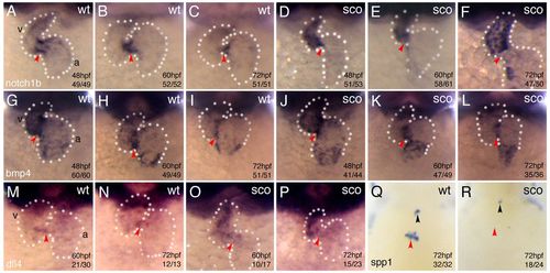
Cardiac valve genes were abnormally expressed in the AVC of scote382 mutant hearts. (A-R) Wild-type (A-C,G-I, M-N,Q) and scote382 (D-F,J?L,O-P,R) embryos were analyzed by in situ hybridization with cardiac valve markers notch1b (A-F), bmp4 (G?L), dll4 (M-P) and spp1 (Q?R) at 48hpf (A,D,G,J), 60hpf (B,E,H,K,M,O), and 72hpf (C,F,I,L,N,P?R). Note that endocardial notch1b and dll4 were ectopically expressed in the ventricle of scote382 mutants but myocardial bmp4 was not affected in scote382 mutants from 48 to 72hpf. spp1, an endocardial?mesenchymal transition marker gene, was not expressed in the AVC of scote382 mutants (red arrowhead in R). The atrium and ventricle are demarcated with red arrowheads. Black arrowheads (Q,R) point to the outflow tract. a, atrium; v, ventricle; wt, wild type. Dotted lines outline the heart. EXPRESSION / LABELING:
PHENOTYPE:
|
|
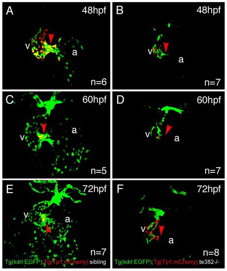
Notch signaling was not activated in the AVC of scote382 mutant hearts. (A?F) Tg(Tp1bglob:hmgb1-mCherry)jh11, called Tg(Tp1:mCherry), transgenic zebrafish (Parsons et al., 2009) were bred with scote382/te382; Tg(kdrl:eGFP) transgenic zebrafish to generate scote382/+; Tg(Tp1:mCherry)/+; Tg(kdrl:eGFP)/+ zebrafish. Images were taken with agarose-embedded embryos using Zeiss 700 upright confocal microscope. Endocardial cells labeled by Tg(kdrl:eGFP) were used to project the heart shapes. Note that Tg(Tp1:mCherry) were gradually restricted in the AVC of wild-type hearts at 48hpf (A), 60hpf (C) to 72hpf (E), but were ectopically expressed in the ventricle of scote382 mutant hearts at 48hpf (B), 60hpf (D) and 72hpf (F). a, atrium; v, ventricle. Red arrowheads point to the AVC. EXPRESSION / LABELING:
PHENOTYPE:
|
|
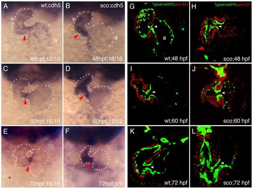
Endocardial cell adhesion and tight junctions were ectopically expressed in the AVC of scote382 mutant hearts. (A?F) Endocardial cell adhesion gene cdh5/ve-cadherin was analyzed by in situ hybridization in wild-type embryos at 48hpf (A), 60hpf (B) and 72hpf (C); and scote382 mutant embryos at 48hpf (D), 60hpf (E) and 72hpf (F). Note gradual restriction of cdh5 in the AVC in wild-type embryos and ectopic expression of cdh5 in the ventricle of scote382 mutant embryos. Red arrowheads point to the AVC. (G?L) Wild-type and scote382 mutant Tg(kdrl:EGFP) transgenic embryos on vibratome sections were subjected to immunostaining with anti-ZO1 antibody. Note that ZO1 was expressed in the AVC of wild-type hearts at 48hpf (G) and downregulated at 60 (I) and 72hpf (K), but remained in the AVC of scote382 mutant hearts at 48 (H), 60 (J) and 72hpf (L). a, atrium; v, ventricle; arrowhead points to the AVC. Dotted lines outline the heart. EXPRESSION / LABELING:
PHENOTYPE:
|
|
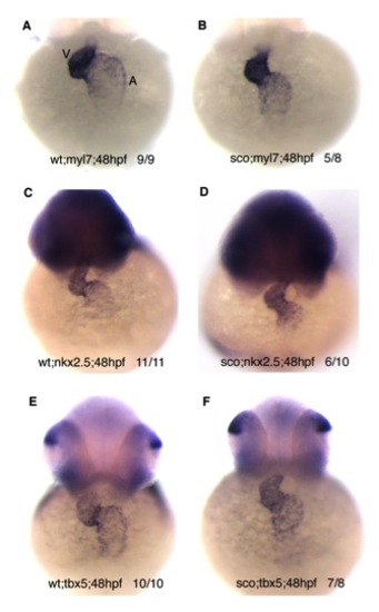
Cardiac genes were not affected in scote382 mutants. Wild-type (A, C, E) and scote382 mutant (B, D, F) embryos at 48 hpf were subjected to whole-mount RNA in situ analyses with myl7 (A, B); nkx2.5 (C, D); or tbx5 (E, F) probes. A, atrium; V, ventricle. |
|
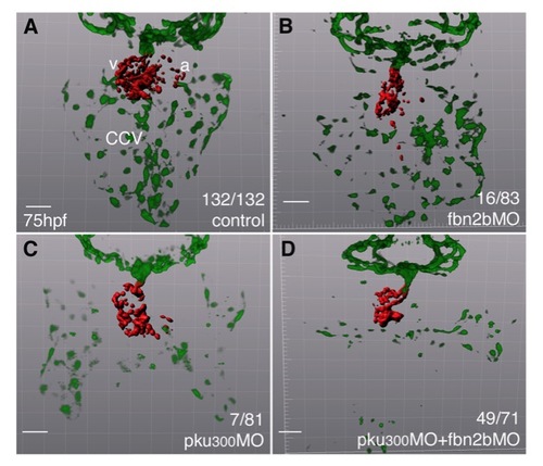
Both fbn2b and pku300 contribute to the formation of atrial endocardium and common cardinal vein (CCV). Compared with wild-type siblings (A), either fbn2bMO (0.4 ng) or pku300MO (4.8 ng) knockdown led to smaller percentages of morphants (16/83 by fbn2bMO; 7/81 by pku300MO) that had defects in the atrial endocardium or CCV (B, C), but simultaneous knockdown by low-dose of fbn2bMO and pku300MO resulted more morphants (49/71) that had defects in the atrial endocardium and CCV (D), suggesting a genetic interaction between fbn2b and pku300 genes. Scale bar, 50 μm. |
|
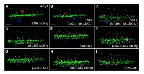
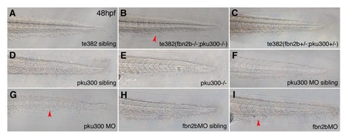
Both fbn2b and pku300 are required for the development of the caudal vein. (A-C) Compared with a wild-type sibling (A), a group II mutant (fbn2b-/-;pku300-/-) had severe defect in the caudal vein (B), while a group I mutant (fbn2b+/-;pku300+/-) had relatively normal caudal vein (C). (D-G) Either a pku300-/- mutant (E) or pku300MO morphant (G) had mild defect in the caudal vein as those of controls (D, F). (H-I) Compared with a fbn2bMO sibling (H), a fbn2bMO morphant had mild defect in the caudal vein (I). Scale bar, 100 μm. |
|
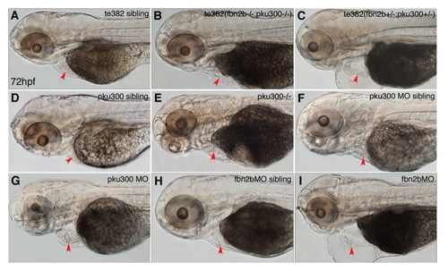
Both fbn2b and pku300 are required for heart development. (A-C) Compared with a wild-type sibling (A), either a group II mutant (fbn2b-/-;pku300 |
|
Both fbn2b and pku300 are required for the development of the ventral tail. (A-C) Compared with a wild-type sibling (A), a group II mutant (fbn2b-/-;pku300-/-) had severe defects in the ventral tail (B), while a group I mutant (fbn2b+/-;pku300+/-) had relatively normal ventral tail (C). (D-G) Either a pku300-/- mutant (E) or pku300MO morphant (G) had mild defects in the ventral tail, compared with those of controls (D, F). (H-I) Compared with a fbn2bMO sibling (H), a fbn2bMO morphant had defects in the ventral tail (I). Red arrowheads point to bubble-like deformed tissues. |
|
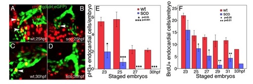
Endocardial cell proliferation is defective in scote382 mutants. (A-E) wild-type (A, C) and scote382 mutant (B, D) Tg(kdrl:eGFP) embryos at 23 hpf and 25 hpf (A, B), 27 hpf and 30 hpf (C, D) were subjected to immunostaining with anti-pH3 antibody. Images were photographed under Zeiss 510 confocal microscope. White arrowheads point to pH3-positive Tg(kdrl:eGFP) endocardial cells in the heart tube. (E) The pH3-positive endocardial cells were scored and statistically analyzed in wild-type and scote382 mutant embryos from 23 to 30 hpf. (F) The BrdU-positive Tg(kdrl:eGFP) endocardial cells were scored and statistically analyzed in wild-type and sco mutant embryos from 23 to 33 hpf. Note that pH3- and BrdU-positive endocardial cells were gradually decreased in scote382 mutants. (E-F) n=3-5; meanąSEM; student?s t-test. |

