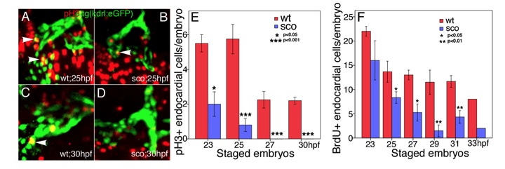
Fig. S6 Endocardial cell proliferation is defective in scote382 mutants. (A-E) wild-type (A, C) and scote382 mutant (B, D) Tg(kdrl:eGFP) embryos at 23 hpf and 25 hpf (A, B), 27 hpf and 30 hpf (C, D) were subjected to immunostaining with anti-pH3 antibody. Images were photographed under Zeiss 510 confocal microscope. White arrowheads point to pH3-positive Tg(kdrl:eGFP) endocardial cells in the heart tube. (E) The pH3-positive endocardial cells were scored and statistically analyzed in wild-type and scote382 mutant embryos from 23 to 30 hpf. (F) The BrdU-positive Tg(kdrl:eGFP) endocardial cells were scored and statistically analyzed in wild-type and sco mutant embryos from 23 to 33 hpf. Note that pH3- and BrdU-positive endocardial cells were gradually decreased in scote382 mutants. (E-F) n=3-5; meanąSEM; student?s t-test.
Image
Figure Caption
Acknowledgments
This image is the copyrighted work of the attributed author or publisher, and
ZFIN has permission only to display this image to its users.
Additional permissions should be obtained from the applicable author or publisher of the image.
Full text @ J. Cell Sci.

