- Title
-
The caudal-related homeobox genes cdx1a and cdx4 act redundantly to regulate hox gene expression and the formation of putative hematopoietic stem cells during zebrafish embryogenesis
- Authors
- Davidson, A.J., and Zon, L.I.
- Source
- Full text @ Dev. Biol.
|
Isolation, mapping, and functional characterization of cdx1a. (A) Predicted peptide alignment of Cdx1 proteins from zebrafish, X. tropicalis, chicken, and human. The sequence of the homeodomain is underlined in red. (B) Genetic and radiation hybrid map positions of cdx1a showing syntenic relationships with human chromosome (Hsa) 5q31–33. (C) Expression of cdx1a (purple) by whole mount in situ hybridization from 50% epiboly (early gastrula) to 48 h post-fertilization (hpf). Embryos in the top panels are oriented with their dorsal side to the right; all others are shown with anterior to the left. Embryos at the 3- and 10-somite stages have been flatmounted and are shown in dorsal views. Transcripts for scl at the 3-somite stage are stained red. Arrowhead indicates expression of cdx1a in the intestinal bulb at 48 hpf (shown in dorsal view). (D) Expression of cdx1a in a control-injected wild-type, cdx1a morphant, cdx4 mutant, and an embryo doubly deficient for cdx1a and cdx4. Arrows indicate the anterior boundary of the cdx1a expression domain in the paraxial mesoderm. (E) Morpholino-mediated knockdown of cdx1a. Images of live embryos injected with cdx1a morpholinos (cdx1amo;cdx4+/− and cdx1amo;cdx4−/−) or mis-match control morpholinos (wild type and cdx4−/−) at 24 hpf. Embryos are shown in lateral views with anterior to the right. Abbreviations: lat., lateral view; veg., vegetal view; tb, tailbud. EXPRESSION / LABELING:
|
|
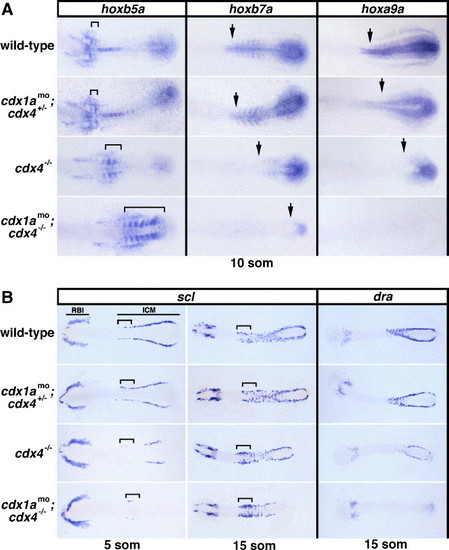
Expression of hox genes, scl, and dra in cdx-deficient embryos. (A) Whole mount in situ hybridizations showing transcripts for hoxb5a, hoxb7a, and hoxa9a in cdx-deficient embryos and wild-type controls at the 10-somite stage. The extent of hoxb5a staining in the somites is indicated with a bracket. Arrows mark the anterior expression boundaries of hoxb7a and hoxa9a in the paraxial mesoderm. Dorsal views are shown, centered on the mid-trunk of flatmounted embryos, with anterior to the left. (B) Expression of scl and dra in cdx-deficient embryos and wild-type controls at the 5- and 15-somite stages. Note the cdx gene dosage effect on scl- and dra-expressing intermediate cell mass (ICM) precursor cells but not on rostral blood island (RBI)-derived cells. Presumptive angioblasts of the rostral ICM are indicated with a bracket and appear expanded in cdx4−/− and cdx1amo;cdx4−/− embryos at the 15-somite stage. Flatmounted embryos are shown in dorsal views with anterior to the left. EXPRESSION / LABELING:
|
|
Expression of flk1 and pax2.1 in cdx-deficient embryos. (A) Whole mount in situ hybridizations showing flk1 transcripts (purple) in cdx-deficient embryos and wild-type controls at the 10- and 15-somite stages (flatmounted embryos shown in dorsal views with anterior to the left) and at 24 hpf (shown in lateral and dorsal views with anterior to the left). Lines mark the rostral angioblast populations. Brackets indicate the regions of flk1-expressing cells near the developing Duct of Cuvier. (B) Whole mount in situ hybridizations showing pax2.1 expression (purple) in the pronephric progenitors (bracket) of wild-type and cdx-deficient embryos at the 10-somite stage. Flatmounted embryos are shown in dorsal views with anterior to the left. EXPRESSION / LABELING:
|
|
Erythropoiesis in cdx-deficient embryos. (A) Whole mount in situ hybridizations showing gata1 transcripts (purple) in cdx-deficient embryos and wild-type controls at the 5-, 10- and 18-somite stages (flatmounted embryos shown in dorsal views of the trunk with anterior to the left) and at 24 hpf (shown in lateral views with anterior to the left). (B) Bar graph showing the number of gata1-expressing cells in cdx-deficient and wild-type embryos at the 5-, 10-, and 18-somite stages. Values represent mean (indicated above each bar) ± SD (n = 4). *Significant difference P < 0.01; **Significant difference P < 0.001 compared to wild-type controls. EXPRESSION / LABELING:
|
|
Rescue of gata1 and runx1a Expression in cdx-deficient Embryos. (A) Whole mount in situ hybridizations showing gata1 transcripts (purple) in wild-type, doubly deficient (cdx1amo;cdx4−/−), hoxb7a-injected, and hoxa9a-injected doubly deficient embryos at the 18-somite stage (flatmounted embryos shown in dorsal views of the trunk with anterior to the left). (B) Expression of runx1a, fli1a, and rag1 in cdx-deficient embryos (lateral views, anterior to the left). Arrows in the runx1a-stained embryos at 36 hpf indicate the extent of artery expression, and higher magnification views of the posterior trunk are shown alongside. A similar region in fli1a-stained embryos is shown with short arrows indicating the dorsal aorta and axial vein. Transcripts for rag1 in the thymus of 4 dpf embryos are indicated by arrowheads. (C) Expression of runx1a in wild-type, cdx4−/− mutants, and hoxa9a-injected cdx4−/− embryos at 48 hpf. Arrows indicate the extent of runx1a staining in the dorsal aorta. Asterisks mark runx1a+ cranial motorneurons in the hindbrain, and the arrowhead in the hoxa9a-rescued embryo indicates the short yolk tube extension. EXPRESSION / LABELING:
|
Reprinted from Developmental Biology, 292(2), Davidson, A.J., and Zon, L.I., The caudal-related homeobox genes cdx1a and cdx4 act redundantly to regulate hox gene expression and the formation of putative hematopoietic stem cells during zebrafish embryogenesis, 506-518, Copyright (2006) with permission from Elsevier. Full text @ Dev. Biol.


