- Title
-
Wnt5 signaling in vertebrate pancreas development
- Authors
- Kim, H.J., Schleiffarth, J.R., Jessurun, J., Sumanas, S., Petryk, A., Lin, S., and Ekker, S.C.
- Source
- Full text @ BMC Biol.
|
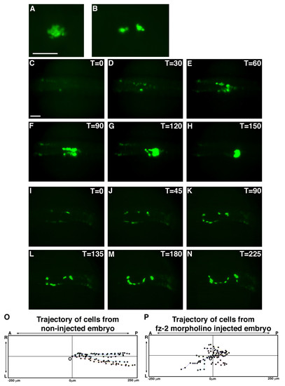
Time-lapse imaging of insulin:GFP transgenic embryos shows cell migration defects in Fz-2 morphants. (A, C-H) Uninjected insulin:GFP transgenic embryo, (B, I-N) fz-2 MO-injected insulin:GFP transgenic embryo. All panels are dorsal views and anterior is to the left. Scale bar represents 100 μm. (A) Uninjected transgenic embryo, 24 hpf. (B) Fz-2 MO-injected transgenic embryo, 24 hpf. (C) At the 14-somite stage, bilateral patches of GFP-positive cells are visible in uninjected embryo. (D) At the 15?16 somite stage, GFP-positive cells have started proliferating. (E-G) At the 17 somite to 24 hpf stages, GFP-positive cells are aligned in bilateral rows of cells and undergo a medial and posterior migration. (H) At 24 hpf, all GFP-positive cells have merged to form one islet. (I) At the 14-somite stage, bilateral patches of GFP expression are apparent in fz-2 MO-injected embryos similar to uninjected embryos. (J-M) GFP-positive cells migrate in random directions in fz-2 morphant embryos. (N) At 24 hpf, GFP-positive cells have still not merged. (O) Trajectory of GFP-positive cells in uninjected insulin:GFP embryo. Notice that cells are uniformly moving posteriorly. (P) Trajectory of GFP-positive cells in fz-2 MO-injected insulin:GFP embryo. Notice cells are moving in random directions. A: anterior, P: posterior, T: time, L: left, R: right, O: origin. |
|
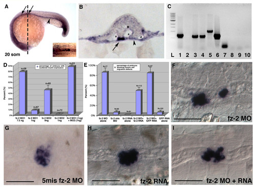
Migration defects in Fz-2 morphant embryos can be rescued by synthetic fz-2 mRNA. (A) Double in situ hybridization with fz-2 and insulin at 20 somite stage of development. Arrow, insulin; arrowhead, fz-2 expression in the endoderm; dotted line, approximate position of the section in (B). (B) A section of double in situ hybridization with fz-2 and insulin. Fz-2 is expressed more strongly on the surface of mesoderm and entire endoderm. Arrow, insulin; arrowhead, fz-2 expression in the endoderm; a, arteries; asterisk, neural tube; d, pronephric duct. (C) RT-PCR using cDNA made from sorted cells of transgenic insulin:GFP zebrafish embryos. L: ladder; lanes 1-5: GFP-negative cells; lanes 6-10: GFP-positive cells; lanes 1, 6: EF1α lanes 2, 7: insulin; lanes 3, 8: fz-2 primer set #1; lanes 4, 9: fz-2 primer set #2; lanes 5, 10: wnt-5. (D) High-dose injection of either fz-2 MO1 or MO2 resulted in scattered insulin expression, whereas low dose injection of either MO caused such defects in less than 10% of embryos. Co-injection of low dose fz-2 MO1 and MO2 resulted in synergistic increase of percentage of embryos with scattered insulin expression. (E) 80% of fz-2 MO-injected embryos displayed scattered insulin expression. Co-injection of fz-2 MO and fz-2 RNA reduced the percentage of embryos with abnormal insulin expression down to 45%. (F-I) In situ hybridization with insulin at 24 hpf stage, anterior is to the left, (F) fz-2 MO1-injected embryo, (G) fz-2 mismatch MO-injected embryo, (H) fz-2 RNA-injected embryo, (I) fz-2 MO- and fz-2 RNA-co-injected embryo. Notice the compact islet in this embryo that displays an undulated notochord. EXPRESSION / LABELING:
PHENOTYPE:
|
|
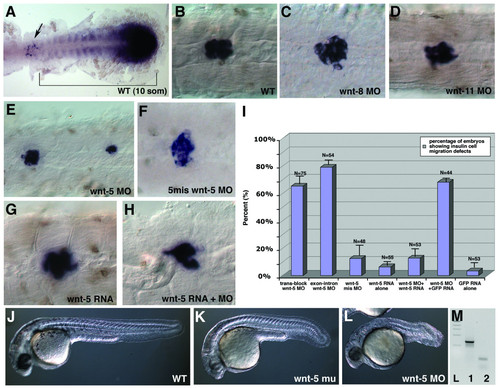
Wnt-5 has a specific role in islet formation. (A) Double in situ hybridization with pdx-1 and wnt-5, 10 som stage, dorsal view, the anterior is to the left. Arrow, pdx-1 expression, bracket, wnt-5 expression. (B-H) In situ hybridization with insulin at 24 hpf. (B) wild-type, (C) WNT-8 morphant embryos, (D) WNT-11 morphant embryos, (E) WNT-5 morphant embryos, (F)wnt-5 mismatch MO-injected embryos, (G) wnt-5 RNA injected embryo, (H) wnt-5 MO and wnt-5 RNA co-injected embryo. Notice the compact islet in this embryo that displays an undulated notochord. (I) Percentage of embryos with scattered insulin expression resulting from injection of wnt-5 MO reduced significantly from 60% to 10% when wnt-5 RNA was co-injected with wnt-5 MO. (J-L) Morphology at 24 hpf, (J) wild-type, (K) wnt-5 insertional mutant, (L) wnt-5 translation-blocking MO-injected embryos. Notice that wnt-5 MO injected embryos have more severe morphological phenotype than wnt-5 insertional mutant embryos. (M) RT-PCR analysis of wnt-5 transcript in wnt-5 exon-intron MO injected embryos. Injection of wnt-5 exon-intron MO results in severely shortened wnt-5 transcript. L:ladder, 1:EF-1 α control, 2:wnt-5. EXPRESSION / LABELING:
PHENOTYPE:
|
|
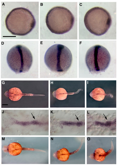
Early endoderm markers are not affected in Wnt-5 and Fz-2 morphant embryos. All pictures are dorsal views. (A, D, G, J, M) wild-type, (B, E, H, K, N) Fz-2 morphants, (C, F, I, L, O) Wnt-5 morphants. (A-C) mixer, 50% epiboly, (D-F) sox-17, 90% epiboly, (G-I) fox-A3, 24 hpf, (J-L) anterior endoderm expression of fox-A3, arrow, pancreatic endoderm, 24 hpf, (M-O) gata-6, 24 hpf. Scale bar = 300 µm. EXPRESSION / LABELING:
|
|
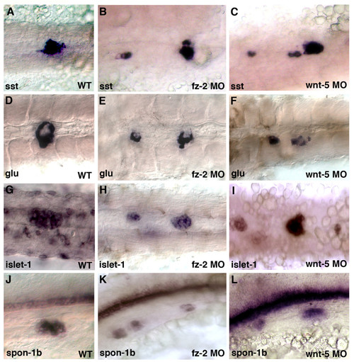
Wnt-5 and Fz-2 morphant embryos exhibit similar pancreatic islet defects at 24 hpf. In all panels, anterior is to the left and 24 hpf. .A-I, dorsal view; J-L, lateral view. (A, D, G, J) Wild-type embryos. (B, E, H, K) Fz-2 morphants. (C, F, I, L) Wnt-5 morphants. In situ hybridization analysis of (A, B, C) somatostatin, (D, E, F) glucagon, notice a hollow spot in the middle of each patch, (G, H, I) islet-1, (J, K, L) fspondin-2b. Note scattered pancreatic cells in Fz-2 and Wnt-5 morphants. |
|
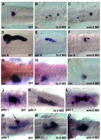
Wnt-5 and Fz-2 morphant embryos have other similar defects. In all panels, view is dorsal, anterior is to the left. (A-I, M-O) 3dpf, (J-L) 24 hpf stage. (A, D, G, J, M) Wild-type embryos. (B, E, H, K, N) Fz-2 morphants. (C, F, I, L, O) Wnt-5 morphants. In situ hybridization analysis of (A-C) insulin, (D-F) carboxypeptidase A, notice the hollow spot indicating the position of the islet, (G-I) ceruloplasmin, (J-O) pdx-1, (M) arrow, pdx-1-staining in islet. EXPRESSION / LABELING:
PHENOTYPE:
|

