- Title
-
Zebrafish foxi1 mediates otic placode formation and jaw development
- Authors
- Solomon, K.S., Kudoh, T., Dawid, I.B., and Fritz, A.
- Source
- Full text @ Development
|
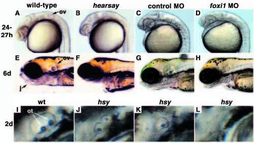
hearsay mutant embryos display defects in otic and jaw development. All panels show lateral views of live embryos with anterior to the left. (A) Wild-type and (B) hsy embryos at 24 hpf. (C,D) Twenty-seven hpf wild-type embryos injected with (C) control morpholino and (D) foxi1 morpholino. (E-H) Six-day-old embryos: (E) wild type, (F) hsy, (G) wild type injected with control morpholino and (H) wild type injected with foxi1 morpholino. (I-L) Otic vesicles in (I) wild-type and (J-L) hsy mutant embryos at 2 days. j, jaw; ot, otolith; ov, otic vesicle. |
|
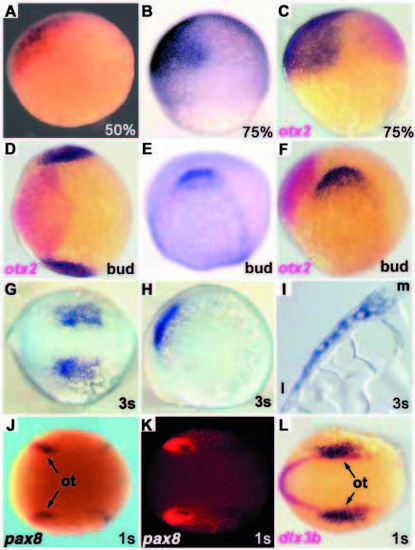
foxi1 expression. All panels show foxi1 expression in dark purple, except J and K (red). Where double in situ labeling is shown, the second marker is indicated in the panel. Anterior is towards the left in D-H,J-O. (A) 50% epiboly and (B,C) 75% epiboly stages, animal pole towards the top, dorsal towards the right. (C) Double in situ labeling with otx2 shown in red. (D-F) Bud stage embryos: (D) dorsal and (E,F) lateral views. (D,F) Double in situ labeling with otx2 in red. (G,H) Three-somite stage embryo: (G) dorsal and (H) lateral views. (I) Transverse section of a three-somite stage embryo. (J-L) One-somite stage embryo: dorsal views. (J,K) Double labeling with pax8 in dark purple, foxi1 in red. (L) double labeling with dlx3b in red. l, lateral; m, medial; ot, otic primordia. EXPRESSION / LABELING:
|
|
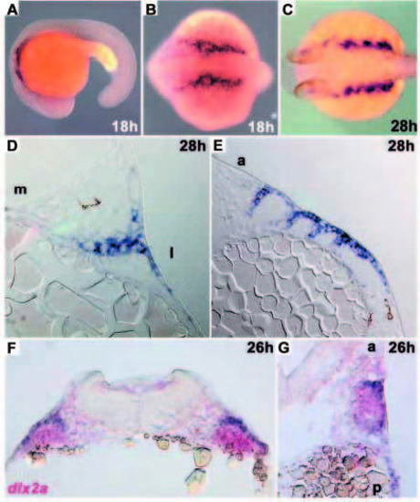
foxi1 pharyngeal arch expression. (A-C) foxi1 expression in whole-mount embryos. (A) 18 hpf, lateral view; (B) 18 hpf, dorsal view; (C) 28 hpf, dorsal view. (D,E) foxi1 expression in 5 μm sections through the pharyngeal arches of 28 hpf embryos. (D) Transverse section and (E) sagittal section. (F,G) 15 μm sections through pharyngeal arch region of 26 hpf embryos: foxi1 expression shown in purple, dlx2a expression shown in red. Transverse (F) and sagittal (G) sections. a, anterior; l, lateral; m, medial; p, posterior. EXPRESSION / LABELING:
|
|
Markers of early placode induction are absent in hsy. (A-F) In situ hybridizations with three- to five-somite stage embryos: dorsal views, anterior towards the left. (A,C,E) wild-type and (B,D,F) hsy embryos. (A,B) pax8 expression, (C,D) pax2a expression and (E,F) dlx3b expression. (G) TaqI genotyping for dlx3b in situ. Migration position for hsy mutant and wild-type fragments are indicated at the left of the gel. mhb, midbrain-hindbrain boundary; ot, otic primordia; pn, pronephros. EXPRESSION / LABELING:
|
|
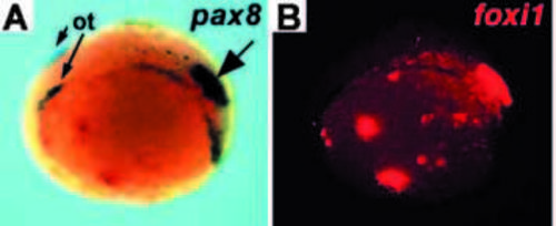
Misexpression of foxi1 can induce ectopic pax8 expression. (A,B) Double in situ labeling of one-somite stage wild-type embryo injected with a foxi1 expression vector. The same embryo is shown in both panels, dorsolateral view, anterior towards the left. (A) pax8 expression shown in dark purple. The large arrow indicates an example of ectopic expression. (B) foxi1 expression shown in red. ot, otic primordia. |
|
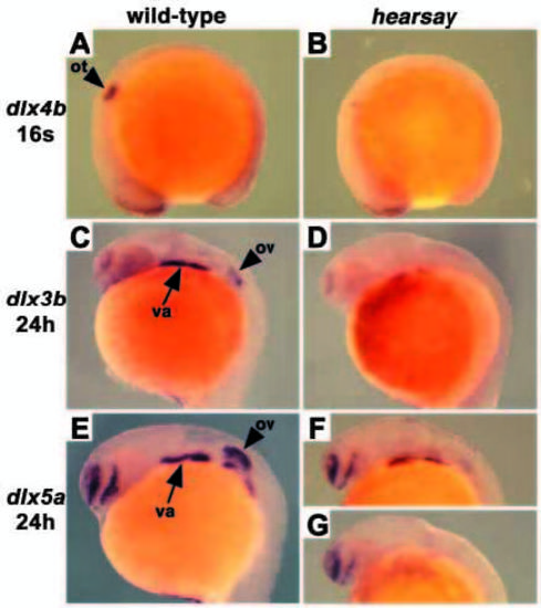
Expression of markers for branchial arches and late-otic development is affected in hsy. RNA in situ hybridization with (A,C,E) wild-type and (B,D,F,G) hsy mutant embryos. (A,B) dlx4b expression, 16-somite stage. (C,D) dlx3b expression, 24 hpf. (E-G) dlx5a expression, 24 hpf. Note the loss of visceral arch expression in G. Forebrain and olfactory placode expression is unaffected in hsy. Lateral views, anterior towards the left in all panels. ot, otic placode; ov, otic vesicle; va, visceral arches. EXPRESSION / LABELING:
|
|
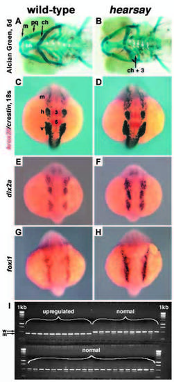
Analysis of jaw and neural crest in hsy. (A,C,E,G) Wild-type and (B,D,F,H) hsy mutant embryos. (A,B) Alcian Green staining of cartilaginous jaw elements at 5 days. Ventral views, anterior towards the left. Cartilages: bh, basihyal; ch, ceratohyal; m, mandibular; pq, palatoquadrate; 3-7, gill cartilages derived from branchial arches P3- P7. m and pq are P1 derivatives; ch and bh are derived from P2. ch+3 indicates unilateral fusion of ceratohyal with gill arch 3 in mutant. (C-H) Expression analysis at the 18-somite stage: dorsal views, anterior towards the top. (C,D) Double labeling with crestin (dark purple) and krox 20 (red). (E,F) dlx2a expression. (G,H) foxi1 expression. (I) TaqI restriction polymorphism genotyping data for foxi1 in situ hybridization at the 18-somite stage, sorted as either ?upregulated? or ?normal expression?. m, mandibular neural crest (nc) stream; h, hyoid nc; v, vagal nc; 3 and 5, krox20 expression in rhombomeres 3 and 5. EXPRESSION / LABELING:
PHENOTYPE:
|

Unillustrated author statements PHENOTYPE:
|

