|
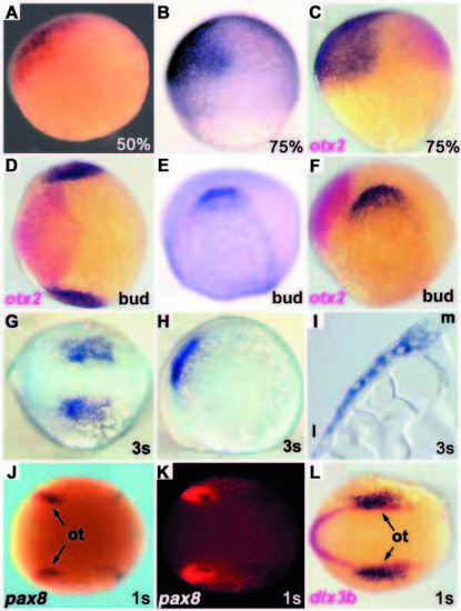
foxi1 expression. All panels show foxi1 expression in dark purple, except J and K (red). Where double in situ labeling is shown, the second marker is indicated in the panel. Anterior is towards the left in D-H,J-O. (A) 50% epiboly and (B,C) 75% epiboly stages, animal pole towards the top, dorsal towards the right. (C) Double in situ labeling with otx2 shown in red. (D-F) Bud stage embryos: (D) dorsal and (E,F) lateral views. (D,F) Double in situ labeling with otx2 in red. (G,H) Three-somite stage embryo: (G) dorsal and (H) lateral views. (I) Transverse section of a three-somite stage embryo. (J-L) One-somite stage embryo: dorsal views. (J,K) Double labeling with pax8 in dark purple, foxi1 in red. (L) double labeling with dlx3b in red. l, lateral; m, medial; ot, otic primordia.
|