- Title
-
Unique and conserved aspects of gut development in zebrafish
- Authors
- Wallace, K.N. and Pack, M.
- Source
- Full text @ Dev. Biol.
|
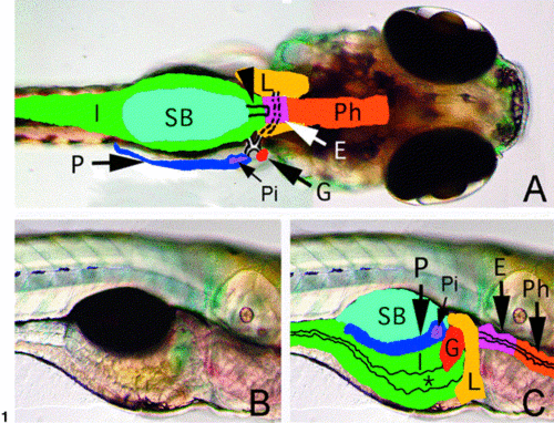
Larval zebrafish digestive system anatomy. (A) Dorsal view of a 5- dpf larva. Overlay outlines the pharynx (Ph), esophagus (E), liver (L), pancreas (P) with solitary islet (Pi), gallbladder (G), swimbladder (SB), and intestine (I). Broken and solid lines depict ducts of the liver, pancreas, and gallbladder. Swimbladder pneumatic duct is denoted by arrowhead. (B, C) Lateral view of 5-dpf larva with overlay. *, marks intestinal lumen. |
|
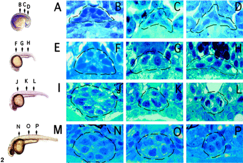
Formation of the zebrafish gut tube at mid-somite stages. (A) Lateral view of an 18-hpf embryo with corresponding histological sections (B?D). Endoderm at this stage does not appear organized. (E) Lateral view of a 21-hpf embryo with corresponding histological sections (F?H). Endoderm near the level of the finbuds (F) assumes a circular or radial organization with peripherally located nuclei. Endoderm caudal to the beginning of the yolk extension (G, H) appears unchanged from 18 hpf. (I) Lateral view of a 26-hpf embryo with corresponding histological sections (J?L). Radially organized endoderm is present rostrally from the finbuds to the yolk extension (J) and near in the common pronephric duct (L) but not within the intervening region (K). (M) Lateral view of a 34-hpf embryo. At this stage, the endoderm from the finbuds to the common pronephric duct has adopted a tube-like configuration. The gut lumen is visible at this stage (N) and also recognizable in transmission electron micrographs of 30-hpf embryos (not shown). Dashed line circles the gut endoderm in all histological sections. |
|
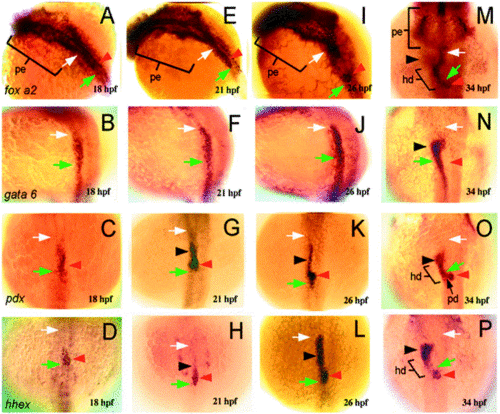
Molecular markers reveal endodermal patterning and organ primordia within the developing zebrafish digestive system. (A?D) 18 hpf. (A) foxA2 expression (dorsolateral view) is restricted to the anterior endoderm, including the pharyngeal endoderm (pe; brackets) and endoderm that lies in between the posterior pharynx (white arrow) and the foregut region (green arrow), which can be identified in histological sections at 21 hpf. (B) gata-6 (dorsolateral view) is expressed within endoderm caudal to the posterior pharynx. gata-6 and foxA2 expression overlap within a small region of endoderm between the posterior pharynx and foregut, whereas endodermal expression of pdx (C; dorsal view) and hhex (D; dorsal view) is restricted to a smaller region of endoderm in this location. (E?H) 21 hpf. At this stage, the expression domains of foxA2 (E) and gata-6 (F) are largely unchanged from 18 hpf. (G) pdx expression is evident within two endodermal domains rostral to the foregut. The caudal region (red arrowhead) is identified as the developing islet, based on insulin immunoreactivity at this location at 24 hpf (not shown). The rostral domain (black arrowhead) is predicted to include progenitors of the liver, and possibly the esophagus and swimbladder, which also arise from endoderm in this region. (H) hhex (red arrowhead) is also expressed within the developing islet. (I?L) 26 hpf. (I) foxA2 expression is strong within the anterior pharynx (pe), posterior pharynx (white arrow), and the pancreatic islet (red arrowhead), but has diminished somewhat within the intervening endoderm. (J) gata-6 expression is largely unchanged. (L) hhex expression in the presumptive liver progenitors (black arrowhead) is pronounced, whereas pdx expression (K) has diminished in this region. At this stage, rostral foregut (green arrow) lies just caudal and ventral to the pancreatic islet. Both pdx and hhex expression persists within the pancreatic islet (red arrowhead) (M?P) 34 hpf. (M) foxA2 expression (dorsal view) persists within the anterior pharynx (pe), posterior pharynx (white arrow), and the pancreatic islet (red arrowhead). Precise identification of intervening endoderm that expresses foxA2 is difficult at this stage but is likely to include cells of the hepatic and pancreatic ducts based on prox-1 immunostainings (not shown). gata-6 (N) expression is now evident in the developing liver (black arrowhead), as is and pdx (O) and hhex (P). pdx expression is also seen within the developing hepatic (hd) and pancreatic ducts (pd). In all panels: white arrow, posterior pharynx; green arrow, foregut; red arrowhead, pancreatic islet; black arrowhead, liver; pe, anterior pharynx; hd, hepatic duct. EXPRESSION / LABELING:
|
|
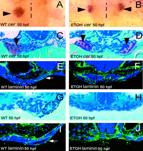
The zebrafish liver and pancreas form independently of the gut tube. (A, B) Whole-mount RNA in situ hybridization using a ceruloplasmin (cer) probe, dorsal view. Compared with WT (A), liver duplication (arrows) is seen in 50-hpf ethanol-treated embryos (B). (C, D) Histological cross-sections through the liver of 50-hpf WT (C) and ethanol-treated (D) embryos processed for cer in situs. Anterior endoderm spans the midline between the duplicated livers (arrows in D) of ethanol-treated embryos. (E, F) Laminin immunostainings from a comparable region of 50-hpf WT (E) and ethanol-treated (F) embryos. Organized endoderm present within the developing esophagus (white arrow) of 50-hpf WT embryos is not identifiable in ethanol-treated embryos (F). (G) Histological cross-sections through the rostral gut of 50-hpf WT embryo. (H) Endoderm in a comparable region of 50-hpf ethanol-treated embryo spans the midline and is unorganized. (I, J) Immunoreactive laminin in a WT 50-hpf embyro (I) encircles the gut tube, but is widely dispersed in unorganized endoderm of ethanol-treated embryos (J). Dotted line in (A, B) represents the embryonic midline. Circle in (G) surrounds the gut tube. |
|
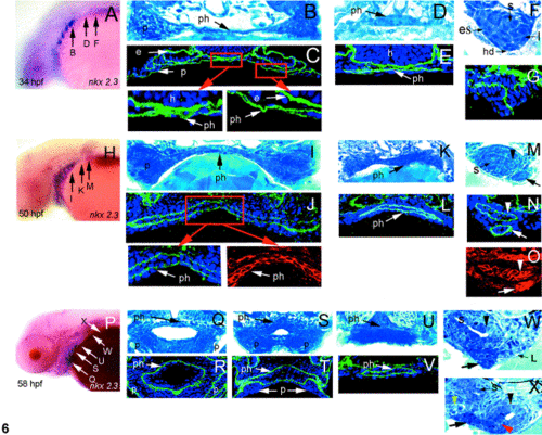
Zebrafish pharynx and esophagus development: morphogenesis and cell polarization. (A?G) 34 hpf. (A) Lateral view of nkx-2.3 expression within the branchial arches with corresponding histological sections (B, D, F). (B) The anterior pharynx is comprised of a single layer of endoderm. Caudally, the endoderm of the posterior (D) pharynx is stratified. (F) Tissue compartmentalization is seen within the region of the developing esophagus. Analysis of caudal serial sections from this and subsequent stages identifies the primordia of the esophagus (e), liver (l), swimbladder (s), and hepatic duct (hd). (C, E, G) 34-hpf laminin immunostainings. At this stage, laminin surrounds individual cells within the anterior and posterior pharynx (C, E) and tissue compartments near the esophageal primordium (G). (H?O) 50 hpf. (H) Lateral view of nkx-2.3 expression. Tissue sections (I) and immunostainings with laminin (green, J) and cadherin (red inset, J) reveal an epithelial bilayer within the anterior pharynx. The posterior pharynx remains stratified (K) and is clearly surrounded by laminin within the developing basement membrane (L). Organization of the esophagus primordium (arrowhead) and swimbladder pneumatic duct (s) is now evident in tissue sections (M) The esophageal epithelium (arrowhead) is now also polarized (N, O). Note also laminin and cadherin in the hepatic duct (arrow). (P?X) 58 hpf. (P) Lateral view of nkx-2.3 expression. (Q) The lumen of the rostral anterior pharynx is visible in tissue sections as are nearby ventral gill slits. (S) The lumen of the anterior pharynx narrows caudally. Note ventral location of the pharyngeal arches in this region (p). (U) The lumen of the posterior pharynx, near the esophageal border, is filled with amorphous debris and difficult to appreciate in tissue sections. (W) At this stage, the esophageal (arrowhead) and swim bladder pneumatic duct (s) lumens are clearly patent. Liver (l) and hepatic duct (arrow) are also seen in this section. Caudally (X), the hepatic?pancreatic duct (red arrowhead) inserts into the developing intestine (black arrowhead). Developing exocrine pancreas (black arrow) and pancreatic islet (yellow arrow) are evident in this section. (y, yolk). (R, T, V) Immunoreactive laminin surrounding the pharynx. In all panels: h, hindbrain; e, ear; p, brancheal arches; ph, pharynx; s, swimbladder; hd, hepatic duct; l, liver; es, esophagus. |
|
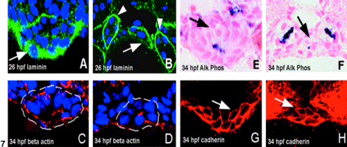
Tissue morphogenesis and cell polarization are coincident within the developing gut tube. (A, B) 26 hpf. Laminin surrounds the newly formed foregut (A, arrow) and hindgut (B, arrow) Arrowheads, pronephric ducts. (C?H) 34 hpf. Apical β-actin (C, D) and alkaline phosphatase activity (E, F) are seen in the gut for the first time at this stage (foregut: white dashed line in C, arrow in E; hindgut: white dashed line in D, arrow in F). Immunoreactive cadherin within the foregut (G arrow) and hindgut (H arrow) is also evident at 34 hpf. |
|
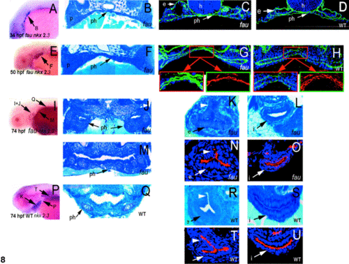
gata-5/fau is required for pharynx morphogenesis but not formation of the esophagus or gut. (A?D) 34 hpf. (A) Lateral view of nkx-2.3 expression within the fau branchial arches with corresponding histological section (B) and laminin immunostaining (C). (B) The fau pharyngeal endoderm appears normal in tissue sections (compare with Fig. 5B). (C) Laminin within the fau pharynx is normal compared with wild-type (D). (E?H) 50 hpf. (E) Lateral view of nkx-2.3 expression within the fau branchial arches with corresponding histological section (F) and laminin (green) and cadherin (red inset) immunostainings in fau (G) and wild-type (H) embryos. The epithelial bilayer within the pharyngeal endoderm that is normally present at 50 hpf (H) is not present in fau mutants (G). (I?U) 74 hpf. (I, P) Lateral view of nkx-2.3 expression within the branchial arches of fau (I) and wild-type (P) larvae with corresponding histological sections (fau: J, M, K, L; and wild-type: Q, R, S). The fau pharynx is duplicated (J; 18%, n = 58) or widened (M) compared with wild-type siblings (Q). In contrast, fau esophagus morphology (K) and differentiation (N) is normal compared with wild-type siblings (R, T). (L) The solitary 74-hpf fau intestine is smaller than that of wild-type siblings (S). Apical binding of a fluorescent lectin (soybean agglutinin) that normally appears after the gut forms is present in both fau (O) and wild-type (U) 74-hpf larvae. In all panels: ph, pharynx; p, branchial arches; e, esophagus; h, hindbrain; I, intestine. EXPRESSION / LABELING:
PHENOTYPE:
|
|
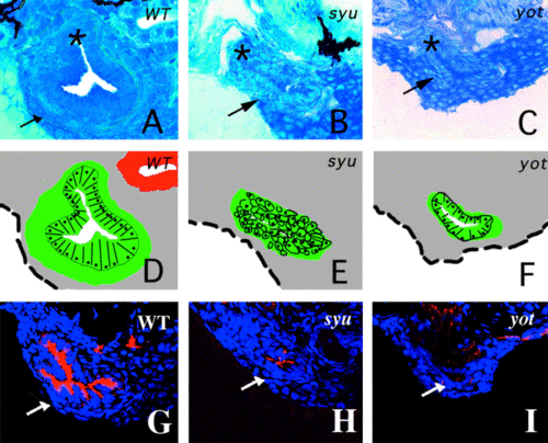
Esophageal development is perturbed by loss of hedgehog signaling. (A?C) Cross-sections through the esophagus of wild-type (A), syu (B), and yto (C) 74-hpf larvae with corresponding schematics (D?F). The wild-type esophagus is comprised of a folded epithelium surrounded by mesenchyme. The swimbladder pneumatic duct (*) branches from the esophagus (arrow) at this level. By contrast, the syu (B) and yto (C) esophagus (arrow) is dysmorphic and binds minimal fluorescent soybean agglutinin (H, I), an epithelial differentiation marker, compared with wild-types (G). PHENOTYPE:
|
|
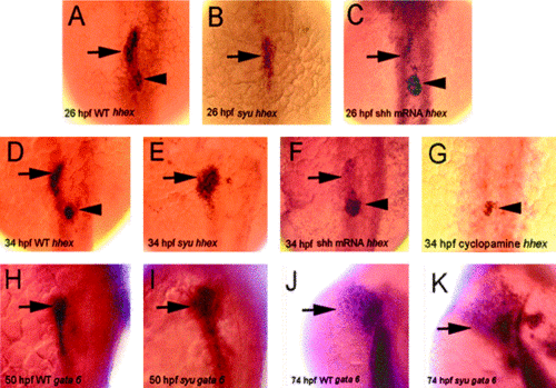
shh regulates zebrafish liver and pancreas development. (A?C) Endodermal hhex expression at 26 hpf, dorsal view. (A) Wild-type liver (arrow) and pancreas (arrowhead) progenitors express hhex. (B) syu mutants lack hhex expression within the developing endocrine pancreas but retain expression within the liver progenitors (arrow). (C, F) Forced expression of shh RNA enlarges the endocrine pancreas (arrowhead) and dramatically reduces the number of liver progenitors (arrow) that express hhex (91% of injected embryos at 26 hpf, n = 47, and 33% of injected embryos at 34 hpf, n = 147). (D, E) hhex expression at 34 hpf, dorsal view. (D) hhex expression within the liver (arrow) and endocrine pancreas (islet, arrowhead) of a wild-type 34-hpf embryo. (E) syu mutants lack hhex expression within the endocrine pancreas and have an enlarged liver (arrow; syu liver enlarged in 65% of 55 random pairs). (G) Embryos treated with cyclopamine at 12 hpf have normal pancreatic hhex expression at 34 hpf (arrowhead), but lack liver progenitors. (H, I) Liver gata-6 expression, 50 hpf (dorsolateral view). The liver of syu mutants (I) is enlarged relative to wild-types (H) (n = 59% of 34 random pairs). (J, K) Liver enlargement in a 74-hpf syu larva (K) compared with a wild-type sibling (J) (n = 20% of 20 random pairs). Liver size comparisons (D, E and G, H and I, J) were made between randomly paired syu mutants and wild-type siblings. At these stages, the syu liver is either enlarged or equal in size to the wild-type liver in all pairs of embryos. EXPRESSION / LABELING:
PHENOTYPE:
|
Reprinted from Developmental Biology, 255(1), Wallace, K.N. and Pack, M., Unique and conserved aspects of gut development in zebrafish, 12-29, Copyright (2003) with permission from Elsevier. Full text @ Dev. Biol.

