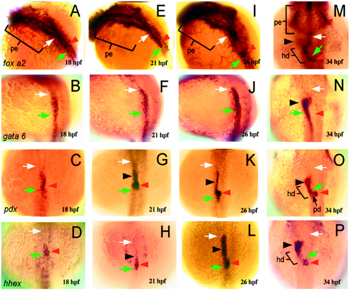
Fig. 3 Molecular markers reveal endodermal patterning and organ primordia within the developing zebrafish digestive system. (A–D) 18 hpf. (A) foxA2 expression (dorsolateral view) is restricted to the anterior endoderm, including the pharyngeal endoderm (pe; brackets) and endoderm that lies in between the posterior pharynx (white arrow) and the foregut region (green arrow), which can be identified in histological sections at 21 hpf. (B) gata-6 (dorsolateral view) is expressed within endoderm caudal to the posterior pharynx. gata-6 and foxA2 expression overlap within a small region of endoderm between the posterior pharynx and foregut, whereas endodermal expression of pdx (C; dorsal view) and hhex (D; dorsal view) is restricted to a smaller region of endoderm in this location. (E–H) 21 hpf. At this stage, the expression domains of foxA2 (E) and gata-6 (F) are largely unchanged from 18 hpf. (G) pdx expression is evident within two endodermal domains rostral to the foregut. The caudal region (red arrowhead) is identified as the developing islet, based on insulin immunoreactivity at this location at 24 hpf (not shown). The rostral domain (black arrowhead) is predicted to include progenitors of the liver, and possibly the esophagus and swimbladder, which also arise from endoderm in this region. (H) hhex (red arrowhead) is also expressed within the developing islet. (I–L) 26 hpf. (I) foxA2 expression is strong within the anterior pharynx (pe), posterior pharynx (white arrow), and the pancreatic islet (red arrowhead), but has diminished somewhat within the intervening endoderm. (J) gata-6 expression is largely unchanged. (L) hhex expression in the presumptive liver progenitors (black arrowhead) is pronounced, whereas pdx expression (K) has diminished in this region. At this stage, rostral foregut (green arrow) lies just caudal and ventral to the pancreatic islet. Both pdx and hhex expression persists within the pancreatic islet (red arrowhead) (M–P) 34 hpf. (M) foxA2 expression (dorsal view) persists within the anterior pharynx (pe), posterior pharynx (white arrow), and the pancreatic islet (red arrowhead). Precise identification of intervening endoderm that expresses foxA2 is difficult at this stage but is likely to include cells of the hepatic and pancreatic ducts based on prox-1 immunostainings (not shown). gata-6 (N) expression is now evident in the developing liver (black arrowhead), as is and pdx (O) and hhex (P). pdx expression is also seen within the developing hepatic (hd) and pancreatic ducts (pd). In all panels: white arrow, posterior pharynx; green arrow, foregut; red arrowhead, pancreatic islet; black arrowhead, liver; pe, anterior pharynx; hd, hepatic duct.
Reprinted from Developmental Biology, 255(1), Wallace, K.N. and Pack, M., Unique and conserved aspects of gut development in zebrafish, 12-29, Copyright (2003) with permission from Elsevier. Full text @ Dev. Biol.

