
Fig. 8
- ID
- ZDB-FIG-240903-169
- Publication
- Wong et al., 2024 - ALS-linked VapB P56S mutation alters neuronal mitochondrial turnover at the synapse
- Other Figures
- All Figure Page
- Back to All Figure Page
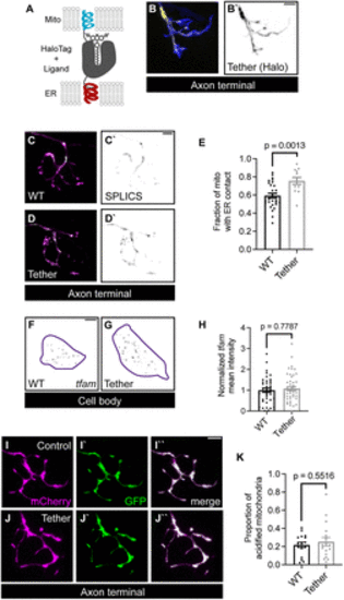
Mitochondrial biogenesis and mitophagy regulation are independent of ER?mitochondrial tethering. A, Schematic of synthetic ER?mitochondrial tether. This tether is composed of the mitochondrial localization sequence from Tomm20 and the ER localization sequence from Sec61? linked with a HaloTag sequence. B, Expression of the synthetic tether (yellow in B, black in B`) in a pLL axon terminal. GFP labels the cytoplasm (blue). C, D, SPLICS signal (green on top, black on bottom) visualizing mitochondria (magenta) proximity to the ER membrane. E, Quantification of SPLICS-positive mitochondrial area in pLL axon terminals. F, G, A representative image of HCR RNA FISH labeled tfam fluorescence in single pLL neuron (purple outline) in individual pLL neurons expressing cell-fill GFP (F) or synthetic tether (G). H, Quantification of mean tfam fluorescence intensity normalized to WT levels. I, J, A representative image of axon terminal GFP and mCherry signals in pLL neuron expressing the mitophagy indicator in WT (I) or the synthetic tether transgenic (J). K, Quantification of the proportion of acidified mitochondria with tether expression. Student's t test for E and K. Mann?Whitney U test for H. Scale bar, 5 ?m. Each data point for E, H, and K represents the average calculated from an individual animal. All data represented as mean � SEM. |