Fig. 6
- ID
- ZDB-FIG-240530-6
- Publication
- Feng et al., 2024 - Core planar cell polarity genes VANGL1 and VANGL2 in predisposition to congenital vertebral malformations
- Other Figures
- All Figure Page
- Back to All Figure Page
|
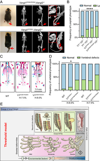
Vangl1-R258H knock-in mice display vertebral defects in a Vangl gene dose- and hypoxia-dependent manner. (A) Phenotype of the 2-mo-old Vangl1R258H/R258H;Vangl2+/? and Vangl1R258H/?;Vangl2+/? mice by MicroCT. Looptail was observed, and vertebral malformations are indicated by red arrows. (B) Penetrance of looptail (Lp) in various Vangl1-R258H mutant mice. The significance of the differences between the groups was calculated by a chi-squared test, ?2 = 27.56, P < 0.0001. (C) Dorsal view of skeletal preparation of E17.5 Vangl1R258H/R258H embryos with hypoxia. Black arrows indicate vertebral ossification abnormalities; blue arrows indicate fused rib; arrowheads indicate hemivertebrae. (D) The penetrance of vertebral defects in wild-type, Vangl1R258H/+, and Vangl1R258H/R258H mouse embryos induced by mild hypoxia. The significance of the differences between the groups was calculated by the chi-squared test, ?2 = 8.00 (H-8.0%), 2.77 (H-7.5%). *P < 0.05. V1 and V2 denote Vangl1 and Vangl2, respectively. H-8.0% and H-7.5% denote hypoxia at 8.0% and 7.5% oxygen concentration, respectively. (E) Threshold model of Vangl gene dosage in birth defects. The decrease in Vangl1 and Vangl2 gene dosage and the addition of environmental factors increase the penetrance and severity of skeletal and neurologic phenotypes. The threshold of Vangl gene dosage for vertebral defects and NT defects is different, with the former more sensitive to Vangl dosage. TSH, tethered spinal cord; DM, diastematomyelia; SM, syringomyelia; SBO, spinal bifida occulta; MC, meningocele; NTDs, neural tube defects. |

