
Fig. 1
- ID
- ZDB-FIG-240530-1
- Publication
- Feng et al., 2024 - Core planar cell polarity genes VANGL1 and VANGL2 in predisposition to congenital vertebral malformations
- Other Figures
- All Figure Page
- Back to All Figure Page
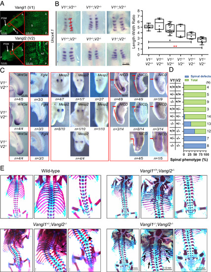
Somite development defects and vertebral malformations in Vangl1 and Vangl2 mutant mouse embryos. (A) Vangl1 and Vangl2 expression in the PSM, NT, and somites (S) of E8.5 wild-type embryos detected by immunofluorescence of endogenous Vangl proteins. A, anterior; P, posterior. (B) Whole-mount in situ hybridizations of Uncx4.1 in 5 to 6 somites stage of mouse embryos. ImageJ quantified the width and length of somites. The width was calculated on the basis of the average of each somite and the length was determined by the Uncx4.1 positive region along the A?P body axis. The significance of the differences between the groups was calculated by a one-way ANOVA test, F = 16.18, *P = 0.0038, **P < 0.0001. Box plots show the center line as the median, box limits as the upper and lower quartiles, and whiskers as the minimum to maximum values. (C) Whole-mount in situ hybridizations of Wnt3a, Fgf4, and Mesp2 and whole-mount immunohistochemistry of NICD in 23 to 25 somite stage of Vangl mutant mice at E9.5. The red arrow and bracket denote a variable cyclic pattern. (D) Penetrance of spinal phenotypes (rib and vertebrae) of Vangl mutant mice. The number of analyzed animals for each genotype is listed on the right side of the chart. (E) Spinal phenotypes of E17.5 to E18.5 Vangl1 and Vangl2 mutant mice. Red asterisks indicate vertebral fusion, vertebral misalignment, and delayed vertebral ossification. Arrowheads point to hemivertebrae, and arrows point to rib fusion, rib shortening/missing, and rib bifurcation. V1 denotes Vangl1, and V2 represents Vangl2. |