Fig. 2
- ID
- ZDB-FIG-240530-2
- Publication
- Feng et al., 2024 - Core planar cell polarity genes VANGL1 and VANGL2 in predisposition to congenital vertebral malformations
- Other Figures
- All Figure Page
- Back to All Figure Page
|
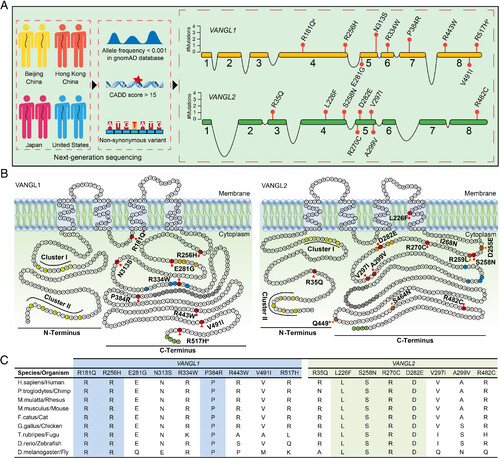
Identification of VANGL1 and VANGL2 variants in CS patients. (A) Brief workflow of the identification of VANGL1 and VANGL2 variants in a multicenter CS cohort (Left) and the location of the identified variants (Right). The numbered orange and green boxes indicate the coding exons of VANGL1 and VANGL2. (B) Amino acid residues of VANGL1 and VANGL2 defining sequence landmarks and signature motifs are depicted in different colors, including rare variants identified in our multicenter cohort (red), looptail variants (dark orange), Curly Bob variant (pink), two highly conserved phosphorylation clusters (yellow, cluster I and cluster II), trans-Golgi network sorting motif (orange), p97/VCP-interacting motif (blue), coiled-coil domain (gray), and PDZ-binding motif (light green). aThe same variant was found in reported NTDs patients. (C) Conservation and multiple alignments of VANGL1 and VANGL2 variants. Highlighted variants are conserved among all listed species. |

